Exploring horizons in 3D microscopy: what the future holds
Posted: 25 July 2018 | Dimitri Scholz (University College Dublin), Karl Gaff (Dublin Institute of Technology) | No comments yet
The microscope slide is flat (2D), but the world around us is not – despite the flat-earth theories. We need volume information about our samples, ideally with high resolution in all three dimensions as well as over time – the fourth dimension…
3D-microscopy is booming; profiting from an increase in computation power. However, improvements in the resolution power of the non-optical in vivo 3D imaging – such as ultrasound tomography, micro-computed tomography (CT), micro-positron emission tomography (PET), photoacoustic imaging and others – will certainly progress,1,2,3 thereby narrowing the gap between these originally 3D, large volume, low-resolution technologies, and the originally high resolution but only 2D microscopy. Classically, samples for microscopy were prepared as thinly as possible to prevent any blurriness from an out-of-focus signal. The standard histological section is 5mm thick,4 which is too thick to be considered flat. Students analysing histological sections realise one could learn more about a sample by simply scrolling though the focusing. Unfortunately, the integrated 3D picture occurs only in the investigator’s brain. So, how best to capture only the in-focus information from each focusing plane and disregard the out-of-focus information? Thankfully, this became possible with motorised focusing enabling precisely stepped acquisition, followed by an appropriate image analysis. For the transmitted and reflected light macro- and microscopy, the Zerene stacker5 is a useful analysis tool for reconstruction of complex surfaces, such as an insect eye or a flower6 (Figure 1).
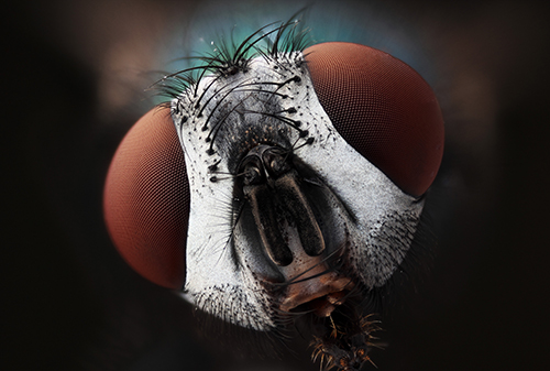
Figure 1. A common green bottle fly (Lucilia sericata) has been imaged using a Canon-MPE 65mm macro photo lens. 85 images with sequential focusing each 23um to cover circa 2mm focus depth were acquired in RAW format with an EOS Canon 60D camera mounted on a motorised focus rail: the maximal focusing projection.
This promising approach is not yet popular in histology.
The higher the numerical aperture7 (NA) of the lens, the thinner the sample must remain strictly in focus in transmission light mode. (The Z-resolution could exceed the XY diffraction-limited resolution, if the 100-200nm sectioning was applied). For NA ≥ 1.4, the sample should be at least 10 times thinner: < 500nm, which is only achievable for samples embedded into a hard resin and sectioned with a diamond or glass knife. This approach was originally developed for transmission electron microscopy. However, this limit of physical sectioning does not apply if the microscope detection system is capable of optical sectioning, such as confocal,8 two-photon,9 light sheet10 or Total internal reflection fluorescence (TIRF) microscopy. TIRF microscopy is intrinsically the 2D application.11 Deconvolution,12,13 developed first for the epi-fluorescent imaging and expanded to all other modes of the fluorescent microscopy,13 such as confocal, spinning disc, two-photon and stimulated emission depletion (STED), provides an additional powerful tool for distilling the in-focus signal. To reveal the 3D information about the sample, one could rotate the sample or the light source, collect multiple images from the different projections, and 3D-reconstruct them using powerful software.
This approach is only recently trying to find its place in light microscopy,14-16 but is broadly used for computer tomography,17 nuclear magnetic resonance tomography18 and transmission electron microscopy.19 A promising and potentially cheaper alternative could be the application of multiple spot-like light sources as described for on-chip holography.20 This could not only reveal the 3D information but also improve the resolution of the imaging system beyond the pixel size. Nobody has tried to combine this with an optical magnification yet. The 3D information can be integrated from multiple physical or optical serial sections. For physical sections, each section must be individually collected and imaged separately. Software corrections must then be carried out for the missing sections, as well as for compression, rotation, tilting, twisting, etc. The entire volume can be then rendered21 in silico.
A remaining challenge for the image analysis is the ratio between the analysed volume and the XYZ-resolution. An XY-scan with a desired magnification can be captured using a motorised stage in a few minutes or even seconds, depending on the number of tiles, and stitched with a FIJI freeware.22 The Z-resolution will be defined by the thickness of the section. The ultimate goal of 3D microscopy is the high resolution in all three dimensions – which is theoretically achievable but remains difficult. In a single experiment, a block of tissue must be serially sliced at ca. 500nm[1] steps for the best possible isometric resolution. (The Z-resolution could exceed the XY diffraction-limited resolution if the 100-200nm sectioning was applied).
For a 1cm3 tissue block, this would require approximately 20,000 sections, each of which must be collected, stained and imaged. Without automation, this would be extremely labour-intensive.
Instead of collecting serial sections, a more promising solution would be the block-face imaging combined with serial block shaving. The term ‘sectioning’ is reserved for the workflow where sections are collected. I introduced ‘shaving’ to emphasise that sections will be discarded.
The clear advantage of this approach is the perfect alignment of serial images: the block does not move. The disadvantage of this approach lies in the difficulty of labelling or staining the entire block of tissue. To overcome this, one can use either auto-fluorescence for imaging or genetically in vivo-introduced fluorescent proteins, which require no further labelling.
Successful 3D reconstructions of a whole mouse brain were demonstrated in 201223 by using a smart combination of green fluorescent protein (GFP) labelling, serial 100um vibratome24 sectioning and serial two-photon imaging. After each vibratome slicing, the block was scanned a number of times at different focal planes deep below the surface, to avoid imaging the layers damaged by the vibratome knife. Only a few vibratome sections were required along with numerous planes of two-photon scanning. With this approach, theoretically the entire tissue could have been imaged with high resolution of about 250-300nm. (Water immersion lenses should be applied to minimise distortions, with maximal NA = 1.2).
However, because the two-photon imaging is a time-consuming process, Ragan23 compromised the Z-resolution for speed but still required several days for an acquisition of the whole mouse brain. The automated cryo-sectioning has been successfully combined with block-face fluorescent imaging.25 Recently, a commercially-available system has been launched, which combines the fluorescence-compatible resin and block-face confocal imaging.26 The advantage of this approach is the possibility to section tissue as thinly as 1mm, which improves Z-resolution. To accelerate imaging, a non-scanned sequential block-face imaging technique must be applied in combination with the sequential ‘shaving’ of the block of tissue with some kind of microtome. The epi-fluorescent microscopy (wide-field) would result in blurred pictures, as the whole block would be illuminated.
Classical confocal microscopy is another scanning approach that shows a comparably slower acquisition rate than two-photon microscopy, and higher blurriness for the block-face imaging. Non-scanning spinning disc confocal microscopy27,28 is fast and suitable for large volume high-resolution imaging of large tissue volumes, but causes even more blurriness in block-face imaging owing to the cross-talking confocal apertures. TIRF29-32 illumination would work well but, unfortunately, the lens-based TIRF requires an oil immersion, which cannot be applied to fresh tissue. For resin-embedded tissue blocks, however, TIRF could become a method of choice for large volume scanning: it is fast, robust and has superb signal-to-noise ratio. Fresh oil must be applied somehow for each XY-scan, but this technology has yet to be built. Light-sheet or single-plane illumination microscopy (SPIM)33 does not require physical sectioning, but the Z-resolution of SPIM is limited by the thickness of the light sheet, which currently cannot be less than several micrometres. Even worse: its unevenness over a larger distance is the factor limiting the image quality and the maximal sample size by only several millimetres. Still, for 4D-microscopy (XYZ + time) of biological samples sized up to several millimetres, SPIM offers the best solution. This technique requires no physical sectioning, but if the tissue is not transparent, it must be fixed and clarified – a technique developed by Werner Spalteholz more than 100 years ago34 and experiencing a renaissance again in contemporary automated 3D microscopy.35,36
The block-face fluorescent imaging is still a great challenge and the answer could be Scanning Electron Microscopy (SEM). The first commercially available automated system for 3D scanning electron microscopy was the Gatan 3View.37 Zeiss has recently brought a similar device to the market. The tissue embedded into a resin block will be shaved by an ultramicrotome and is then block-face imaged by a scanning electron microscope. The XY-resolution can be as high as 5nm, Z-resolution depends on section thickness and can be as high as 30nm (usually 50-100nm); and the volume can be as large as 1mm3. In comparison, the focused ion beam scanning electron microscopes (FIB-SEM)38 (3D microscopes) can reconstruct information from maximal 20-50mm in depth, and TEM tomography can only cover less than 300nm in depth (however with a better XYZ resolution). The Gatan 3View user must compromise between the sample volume and the resolution for the sake of time and data load.
The 3D-SEM technologies hold great promise for future research.38 They will undoubtedly evolve towards the reconstruction of ever-larger volumes, thereby competing with light microscopy-based methods. From a medical perspective, 3D imaging and reconstruction of centimetre-scale blocks (eg, imaging of the entire solid tumour) would open completely new horizons. A low-power SEM provides a higher resolution and better structural recognition than light microscopy. A non-expensive table-top SEM, combined with an automated microtome, would make an automated 3D microscope for larger pieces of tissue up to several cm3. This system could possess the XYZ-resolution of 100nm and cover a large part of biomedical applications in the future. The tissue can be embedded into soft wax, rather than hard resin, and sectioned with a razor blade-microtome. The block could be etched with chloroform vapours before each sequential imaging to reveal the structures and antigens for labelling.
Biographies
 DR DIMITRI SCHOLZ completed his PhD at the National Medical University, Moscow, Russia, his Habilitation (Dr of Sciences) at Goethe University Frankfurt/ Main, Germany, for cell biology and microscopical anatomy. After academic positions at Max-Planck Institute for Experimental Cardiology in Bad Nauheim (Germany) and at Medical University of South Carolina (MUSC, Charleston SC, USA), he joined UCD in Dublin (Ireland) as the founder and head of the Imaging facility. His research interests belong to pushing frontiers of microscopy for cell biology, developing new microscopical tools, systems and approaches.
KARL GAFF is a Laboratory Technician at Dublin Institute of Technology.
References
- du Plessis A, Broeckhoven C, Guelpa A, le Roux S. Laboratory x-ray micro-computed tomography: a user guideline for biological samples. GigaScience. 2017;6:1-11.
- Gauberti M, Fournier A, Docagne F, Vivien D, de Lizarrondo S. Molecular Magnetic Resonance Imaging of Endothelial Activation in the Central Nervous System. Theranostics. 2018;8(5):1195-212.
- de la Zerda A, Liu Z, Zavaleta C, Bodapati S, Teed R, Vaithilinga S, et al. Enhanced Sensitivity Carbon Nanotubes as Targeted Photoacoustic Molecular Imaging Agents. In: Oraevsky A, LV W, editors. Proc of SPIE Vol 7177. 71772009.
- M. M, U. W. Romeis – Mikroskopische Technik: Springer Spektrum; 2015.
- Zerene. Zerene Stacker – The Basics [Available from: http://zerenesystems.com/cms/stacker.
- Gaff K. Optical Botany [Available from: https://www.flickr.com/photos/38484518@N07/albums/72157662995635935.]
- Zeiss-Campus. Numerical Aperture and Light Cone Geometry [Available from: http://zeiss-campus.magnet.fsu.edu/mobile/tutorials/basics/numericalaperturelightcones/indexmobile.html.
- Pawley J. Handbook of biological confocal microscopy: Springer; 2006.
- Helmchen F, Denk W. Deep tissue two-photon microscopy. NATURE METHODS. 2005;2:932-40.
- Elisa Z, Toon B, De Smedt S, Katrien R, Kristiaan N, Kevin B. Technical implementations of light sheet microscopy. Microsc Res Tech. 2018.
- Poulter N, Pitkeathly W, Smith P, Rappoport J. The physical basis of total internal reflection fluorescence (TIRF) microscopy and its cellular applications. Methods Mol Biol. 2015;1251:1-23.
- SVI. Huygens deconvolution [Available from: https://svi.nl/HuygensDeconvolution].
- Cybernetics M. AutoQuant Deconvolution Algorithms [Available from: http://www.mediacy.com/index.aspx?page=Auto_Algorithm].
- Charrière F, Marian A, Montfort F, Kuehn J. Colomb T. Cell refractive index tomography by digital holographic microscopy. OPTICS LETTERS. 2006;31(2):178-80.
- Bruns T, Schickinger S, Schneckenburger H. Sample holder for axial rotation of specimens in 3D microscopy. Journal of Microscopy. 2015;260(1):30-6.
- Lanzano L, Hernandez I, Castello M, Gratton E, Diaspro A, Vicidomini G. Encoding and decoding spatio-temporal information for super-resolution microscopy. Nature Communications. 2015;6:6701-9.
- FDA. What is Computed Tomography? [Available from: http://www.fda.gov/Radiation-EmittingProducts/RadiationEmittingProductsandProcedures/MedicalImaging/MedicalX-Rays/ucm115318.htm.
- Klioze. Basic Physics & a Brief History 2013 [Available from: https://www.youtube.com/watch?v=djAxjtN_7VE].
- Schröder R. Advances in electron microscopy: A qualitative view of instrumentation development for macromolecular imaging and tomography. Archives of Biochemistry and Biophysics. 2015; 581:25–38.
- Göröcs Z, Ozcan A. On-Chip Biomedical Imaging. IEEE Rev Biomed Eng. 2013;6:29–46.
- Bitplane. Imaris [Available from: http://www.bitplane.com/imaris/imaris.
- NIH. Fiji Is Just Image J 2018 [Available from: https://imagej.net/Fiji.
- Ragan T, Kadiri L, Venkataraju K, Bahlmann K, Sutin J, Taranda J, et al. Serial two-photon tomography: an automated method for ex-vivo mouse brain imaging. Nat Methods. 2012;9(3):255-8.
- Abdelaal H, Kim H, Wagstaff R, Sawahata R, Southern P, Skinner P. Comparison of Vibratome and Compresstome sectioning of fresh primate lymphoid and genital tissues for in situ MHC-tetramer and immunofluorescence staining. Biological Procedures Online. 2015;17:2-7.
- Roy D, Steyer G, Gargesha M, Stone M, Wilson D. 3D Cryo-Imaging: A Very High-Resolution View of the Whole Mouse. Anat Rec. 2009;292(3):342–51.
- Hoehn K, Fuchs J, Froeber A, Kirmse R, Glass B, Anders-Oesswein M, et al. Preservation of protein fluorescence in embedded human dendritic cells for targeted 3D light and electron microscopy. Journal of Microscopy. 2015;259(2):121–8.
- Andor. The Revolution WD and XD [Available from: http://www.andor.com/microscopy-systems/revolution].
- Zeiss. Introduction to Spinning Disc Confocal microscopy [Available from: http://zeiss-campus.magnet.fsu.edu/articles/spinningdisk/introduction.html].
- Zeiss-Campus. Education in Microscopy and Digital Imaging 2015 [Available from: http://zeiss-campus.magnet.fsu.edu.
- Microsystems L. Science Lab 2015 [Available from: http://www.leica-microsystems.com/science-lab/topics/.
- Olympus. Microscope resource center 2015 [Available from: http://www.olympusmicro.com].
- Nikon. Nikon MicroscopyU – The Source for Microscopy Education 2015 [Available from: http://www.microscopyu.com].
- Pitrone P, Schindelin J, Stuyvenberg L, Preibisch S, Weber M, Eliceiri K, et al. OpenSPIM: an open-access light-sheet microscopy platform. NATURE METHODS. 2013;10:598–9.
- Spalteholz W. Mikroskopie und Mikrochemie Betrachtungen über die Grundlagen der mikroskopischen Untersuchungsmethoden. Leipzig Hirzel; 1904.
- Tomer R, Ye L, Hsueh B, Deisseroth K. Advanced CLARITY for rapid and high-resolution imaging of intact tissues. Nature Protocols. 2014;9:1682–97.
- Marx V. Optimizing probes to image cleared tissue. Nature Methods. 2016;13(3):205-9.
- Gatan. 3View System [Available from: http://www.gatan.com/products/sem-imaging-spectroscopy/3view-system.
- Zeiss. FIB-SEMs. Focused Ion Beam Scanning Electron Microscopes [Available from: http://www.zeiss.com/microscopy/en_de/products/fib-sem-instruments.html.
- Hughes L, Hawes C, Monteith S, Vaughan S. Serial block face scanning electron microscopy – the future of cell ultrastructure imaging. Protoplasma 2014 Mar;251(2):. 2014;251(2):395-401.
The rest of this content is restricted - login or subscribe free to access
 Thank you for visiting our website. To access this content in full you'll need to login. It's completely free to subscribe, and in less than a minute you can continue reading. If you've already subscribed, great - just login.
Why subscribe? Join our growing community of thousands of industry professionals and gain access to:
- quarterly issues in print and/or digital format
- case studies, whitepapers, webinars and industry-leading content
- breaking news and features
- our extensive online archive of thousands of articles and years of past issues
- ...And it's all free!
Click here to Subscribe today Login here
Related topics
Computerised Tomography (CT), Imaging, In Vivo, Microscopy, Positron emission tomography (PET)