Applications of high content screening in autophagy
Posted: 4 September 2018 | Ole Pless, Saba Ezazi Erdi, Sheraz Gul | No comments yet
Autophagy is an important process to maintain cellular homeostasis and function.1 Basal levels of autophagy are essential for most cells to remove unwanted protein aggregates and damaged organelles in order to prevent diseases.2 However, sometimes cells are unable to maintain physiological stability as a consequence of altered autophagy, which leads to various pathological conditions, such as cancer, cardiomyopathies or neurodegenerative diseases. The successful manipulation of autophagy could revolutionise the way we treat these conditions.
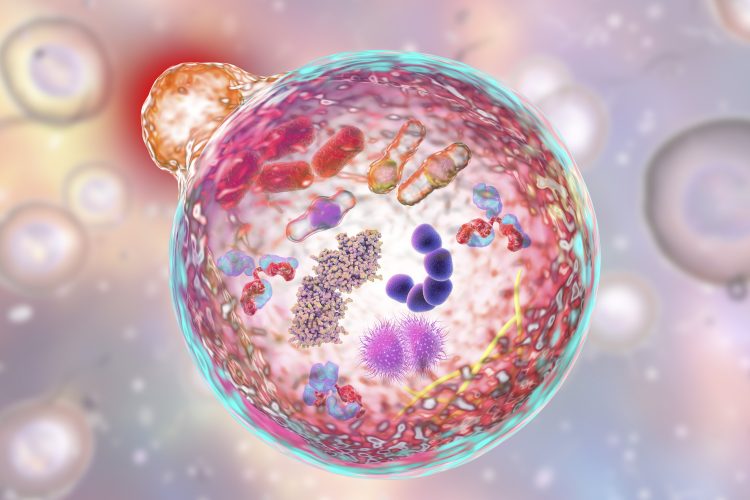

In 2016, The Nobel Prize in Physiology or Medicine was awarded to a molecular biologist, Yoshinori Ohsumi, for his work in the field of autophagy and discovery of its underlying mechanism.3 This work led to the identification of genes essential for autophagy in yeast and explanation of the processes behind it.4 This critical work enhanced our understanding of the human body and emphasised the importance of autophagy in many biological processes and diseases; it also drew the attention of many scientists in both academia and industry to the subject. Autophagy, as well as other complex cellular processes, can be analysed using sophisticated automated microscope-based screening technology – ie, High Content Screening (HCS) – and the use of this technique has grown significantly in recent years due to its versatility in enabling the study of many cellular features and processes simultaneously in complex biological systems. This ability to multiplex many features is what gives HCS remarkable power in drug discovery as it allows the measurement of any biological activity within cells or whole organisms. It also has the capability to identify and validate new drug targets, enable the understanding of the mechanism of action of compounds that are starting points for drugs, predict in vivo toxicity, and propose pathways or molecular targets of orphan compounds. HCS can be used at various stages of the drug discovery pipeline – even to support clinical trials.5
Traditionally, lysosome detection was used as an indicator for autophagy and was measured by electron microscopy or biochemical assays.4 The number and activity of lysosomes, however, turned out not to be a reliable measure of autophagy and new assays that can more accurately assess autophagic activity emerged.6 Yeast has turned out to be the ideal organism with which to study autophagy genes, as within just a few hours of nutrient starvation autophagic bodies can be readily detected. This finding enabled the genetic screening of mutants that are deficient for starvation-induced autophagy pathways and led to the identification of autophagy-related genes (ATG). The knowledge obtained from yeast also facilitated subsequent research in mammals and plants,4 since most ATG genes participating in yeast autophagy were found to have mammalian homologues, and some also with homologues in plants.7
In mammals, regulation of autophagy is more complex than in yeast. Depletion of total amino acids strongly induces autophagy in many types of cultured cells, but the effects of individual amino acid depletion differ based on the type of cells analysed. Amino acids not only have an intermediary role in metabolism but also stimulate target of rapamycin (MTOR)-mediated signal transduction, which controls many pathways, including autophagy.8 Other important key regulators of autophagy are the Beclin 1 complex, the class III phosphatidylinositol 3-kinase (PI3KC3) and the microtubule-associated protein 1 light-chain 3 (LC3).2 Research in the field of autophagy is rapidly evolving and, within the past decade, numerous assays have been developed to monitor autophagy at different stages of the process. Technique selection depends on what the researcher wants to measure: number of autophagosomes, autophagic flux, and activation or inhibition of the pathway.4 There is no single perfect method available to measure all aspects of autophagy. Instead, a combination of techniques should be considered to accurately assess autophagic activity in any biological setting to reduce limitations. One way of finding the right approach for drug discovery is choosing the most appropriate technology platform.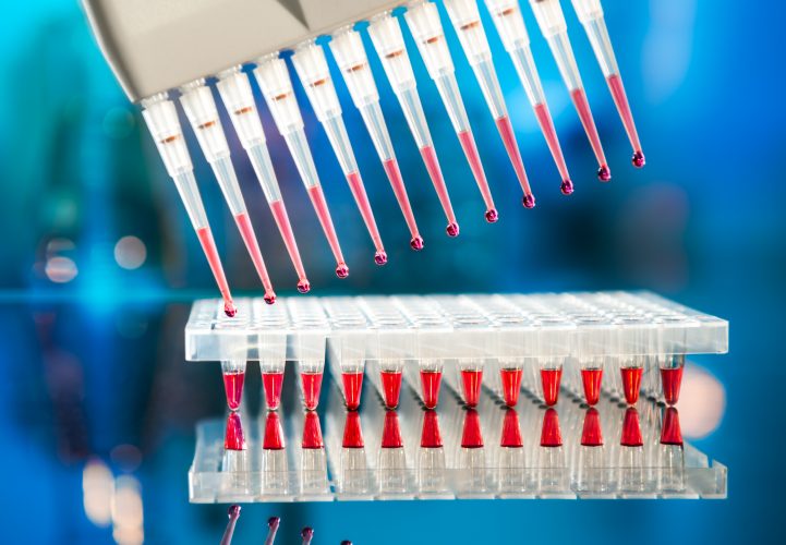

Several drugs approved by the Food and Drug Administration (FDA) modulate autophagy signalling, suggesting that modulation of autophagy with pharmacological agonists or antagonists provides a potential therapeutic approach to target autophagy-related diseases. Thus, specific small molecule modulators of autophagy could serve as tools for studying autophagy-related pathways or even modulate disease-associated phenotypes as a future therapeutic intervention strategy. A particularly interesting high-content assay to measure the autophagic flux within cells, which is commonly used these days, is the GFP-RFP-LC3 dual-reporter assay. LC3 has many functions in autophagy and thus can serve as a specific biomarker for this process. During autophagosome synthesis, the cytoplasmic localisation of microtubule-associated protein 1 light chain 3-I (LC3-I) is conjugated with phosphatidylethanolamine (PE) and becomes the membrane-bound form LC3-II, which is in both the outer and inner membranes of the autophagosomes. Due to this property, analysis of the colour change in fluorescent-tagged LC3 is a widely used technique for LC3 localisation and identification of autophagosomes.9 Potential hits from a cellular or biochemical assay can then be confirmed in their activity in physiologically more relevant secondary assays; eg, by using human induced pluripotent stem cell- (hiPSC) based models. hiPSCs could potentially revolutionise drug discovery since they more faithfully reflect physiological and pathophysiological conditions in vitro compared to ‘classic’ transformed cell lines.10 In addition, they can be expanded to theoretically unlimited numbers of genetically ‘normal’ cell material for high throughput screening purposes. Furthermore, many somatic cell types of the human body have already been generated from hiPSC, including cardiac, neuronal and endodermal secretory cell types. Using the advanced knowledge available in generating and quality controlling hiPSC lines, it is possible to apply hiPSC technology to study autophagy mechanisms in potentially all human cell types. With the appropriate tools and technology, as well as cellular models, autophagy assays can be miniaturised for HTS with the ultimate goal of processing larger compound sets and validating drug candidates for preclinical testing in a timely and efficient manner.
Once bona fide modulators of autophagy have been identified and validated in orthogonal assays, it is essential to attribute any compound-mediated change in cellular responses to autophagy and rule out other effects such as cytotoxicity, apoptosis, etc.11 In addition, if these compounds are to progress in the drug discovery value chain, they should not be associated with significant undesirable characteristics that could lead to a safety issue in the clinic.12-15 Therefore, identifying potential problems with a compound as early as possible will de-risk the project and increase the chances of it successfully progressing in the right direction. It is therefore important to understand compound-mediated cell toxicity, which is often based on the integrity of the cell membrane, key cellular biochemical reactions, and specific cellular markers. Quantification of the extent to which these are compromised allows the viability of the cells to be assessed. A variety of microtitre plate-based cytotoxicity assays based on colourimetric, fluorometric and luminescence detection technologies are available. The absorbance-based methods have historically been the most widely utilised and are well validated. These include quantification of (1) mitochondrial succinate dehydrogenase activity of cells using the tetrazoles XTI and MTT, (2) extracellular lactate dehydrogenase activity by measuring NADH consumption, (3) acid phosphatase activity as a marker of lysosomal activity using p-nitrophenyl-phosphate, (4) remaining glucose in cell culture medium using a glucose oxidase-peroxidase assay, (5) cell proliferation using crystal violet dye accumulation in the nucleus, (6) lysosomal accumulation of the cationic dye neutral red and (7) protein synthesis using sulforhodamine B binding to proteins.
Fluorometric-based cytotoxicity assays are also available and include those that quantify (1) lactate dehydrogenase activity assays using a coupled reaction that results in the conversion of resazurin into resorufin (CytoTox-ONE Assay), (2) protease activity that is released from cells with a compromised cell membrane and a non-cell permeable fluorogenic peptide substrate (CytoTox-Fiuor Cytotoxicity Assay) and (3) the activity of two proteases using cell permeable (giving the live cell measurement) and non-cell permeable (giving the dead cell measurement) substrates (MultiTox-Fluor Multiplex Cytotoxicity Assay).
Luminescence-based assays are also available and include those that quantify (1) the activity of a protease that is released from cells that no longer retain an intact cell membrane using an luminogenic peptide substrate (CytoTox-Glo Cytotoxicity Assay), (2) glutathione-5-transferase activity using a luciferin-derived substrate, the product of the reaction being a substrate of firefly luciferase, and (3) intracellular ATP using a luciferase reaction (CellTiter-Glo Luminescent Cell Viability Assay). There is also a multiplex assay that uses a fluorescence and luminescence readout: essentially the same as the MultiTox-Fluor Multiplex Cytotoxicity Assay mentioned above, but the substrate for dead cells is luminogenic instead of being fluorogenic.
As cell-based assays are now becoming a preferred format for screening, it is necessary to ascertain that the compounds identified are genuine mediators of the activity of the target of interest. This may in part be determined using a suitable target-based secondary assay where the activity of the target can be monitored in an independent assay format. However, this assay itself may be a cell-based one and the question about compound-induced cytotoxicity remains, which can lead to artefacts and complicate compound progression strategies.
Biography


References
- Jing K, Lim K. Why is autophagy important in human diseases? Mol. Med. 2012, 44, 69–72.
- Peppard JV, Rugg C, Smicker M, Dureuil C, Ronan B, Flamand O, Durand L, Pasquier B. Identifying Small Molecules which Inhibit Autophagy: a Phenotypic Screen Using Image-Based High-Content Cell Analysis. Chem. Gen. Trans. Med. 2014, 8, (Suppl-1, M2) 3–15.
- Tooze SA, Dikic I. Autophagy Captures the Nobel Prize. Cell. 2016, 167, 1433–1435.
- Nakatogawa H, Suzuki K, Kamada Y, Ohsumi Y. Dynamics and diversity in autophagy mechanisms: lessons from yeast. Rev. Mol. Cell Bio. 2009, 458–467.
- Buchser W, Collins M, Garyantes T, Guha R, Haney S, Lemmon V, Li Z, Trask OJ. Assay Development Guidelines for Image-Based High Content Screening, High Content Analysis and High Content Imaging. Assay Guidance Manual [https://www.ncbi.nlm.nih.gov/books/NBK100913/].
- Mizushima N, Yoshimorim T, Levine B. Methods in Mammalian Autophagy Research. Cell. 2010, 140, 313–326.
- Díaz-Troya S, Pérez-Pérez ME, Florencio FJ, Crespo JL. The role of TOR in autophagy regulation from yeast to plants and mammals. Autophagy. 2008 7, 851–65.
- Meijer AJ, Lorin S, Blommaart EF, Codogno P. Regulation of autophagy by amino acids and MTOR-dependent signal transduction. Amino Acids, 2015, 47, 2037–2063.
- Zhou C, Zhong W, Zhou J, Sheng F, Fang Z, Wei Y, Chen Y, Deng X, Xia B, Lin J. Monitoring autophagic flux by an improved tandem fluorescent-tagged LC3 (mTagRFP-mWasabi-LC3) reveals that high-dose rapamycin impairs autophagic flux in cancer cells. Autophagy, 2012, 8, 1215–
- Jungverdorben J, Till A, Brüstle O. Induced pluripotent stem cell-based modeling of neurodegenerative diseases: a focus on autophagy. Mol. Med. 2017, 95, 705–718.
- An WF, Tolliday N. Cell-based assays for high-throughput screening. Biotechnol. 2010, 45, 180–186.
- Mandavilli BS, Aggeler RJ, Chambers KM. Tools to Measure Cell Health and Cytotoxicity Using High Content Imaging and Analysis. Mol. Biol. 2018, 1683, 33-46.
- Méry B, Guy JB, Vallard A, Espenel S, Ardail D, Rodriguez-Lafrasse C, Rancoule C, Magné N. In Vitro Cell Death Determination for Drug Discovery: A Landscape Review of Real Issues. Cell Death. 2017, 10:1179670717691251.
- Adan A, Kiraz Y, Baran Y. Cell Proliferation and Cytotoxicity Assays. Pharm. Biotechnol. 2016, 14, 1213–1221.
- Parboosing R, Mzobe G, Chonco L, Moodley I. Cell-based Assays for Assessing Toxicity: A Basic Guide. Chem. 2016, 13, 13–21.
The rest of this content is restricted - login or subscribe free to access

Why subscribe? Join our growing community of thousands of industry professionals and gain access to:
- quarterly issues in print and/or digital format
- case studies, whitepapers, webinars and industry-leading content
- breaking news and features
- our extensive online archive of thousands of articles and years of past issues
- ...And it's all free!
Click here to Subscribe today Login here
Related topics
Assays, Drug Development, Drug Discovery
Related organisations
Fraunhofer-IME SP


