Israeli researchers develop new model that will help pinpoint complications in individuals with Differences in sex development (DSD).
Researchers, led by Dr Nitzan Gonen, of Bar-Ilan University's Goodman Faculty of Life Sciences and Institute of Nanotechnology and Advanced Materials, Israel, offered a solution to one the significant challenges in differences in sex development (DSD), that is the lack of an in vitro system to model and study variants found in these individuals. The study was recently published in Science Advances.
According to the UK National Health Services (NHS), DSD is a group of rare conditions involving genes, hormones and reproductive organs, including genitals. It means a person's sex development is different to most other people's
One of the significant challenges in DSD research is the lack of an in vitro system to model and study variants found in DSD individuals. Through the development of new tools, the researchers, created the somatic/supporting cells of the gonad and were able to model stem cells derived from a DSD individual in a dish. This facilitated the ability to start investigating DSD pathologies in the lab dish in a human-related context.
Stem cells can be used to make both egg progenitor cells and ovarian somatic cells. When the researchers combined these two cells together, they were able to create a healthy and fertile embryo as a product of these artificial eggs.
Male testicles contain germ cells that develop into sperm cells starting from puberty and throughout life. These cells are surrounded by supporting somatic (non-germ) cells that allow germ cells to function and develop.
The team differentiated mouse embryonic stem cells into early somatic cells in the testis. They compared the cells they created to real testis cells and showed, through RNA sequencing technology, that the two are very similar.
The advantage of using mouse-derived stem cells first is that it enables proper comparison to real gonadal cells, isolated from embryonic gonads. It is very difficult, even impossible, to do this with human cells and human embryos, because the human equivalent is an embryo at week seven of pregnancy, a stage where access to embryos following abortions is challenging.
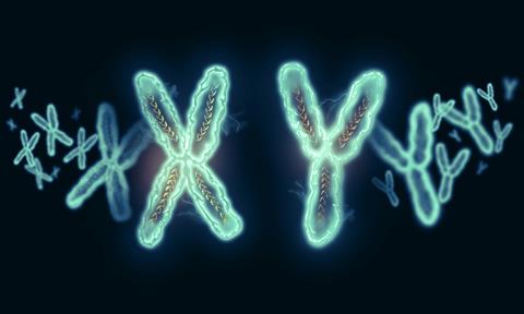
The researchers took three types of human cells: XY, XX and cells from a DSD individual (XY born as female). They showed that the somatic cells produced from XX and XY are different from each other, while the cells of the DSD individual are somewhere in the middle, closer to a female than to a male. However, when the variant in the DSD individual cells was corrected with CRISPR genome editing technology, the cells returned to behaving like typical XY cells.
The creation of this cellular model for a human with DSD opens the door to understanding why the chromosomal patterns differ from XX or XY and what exactly is altered in the DSD individual’s cells.
“Being able to understand the reasons behind differences in sex development is often very valuable to the individual affected and to their family. Additionally, it is often important when deciding on possible clinical treatments. For example, to potentially preserve, maintain or restore fertility," said Robin Lovell-Badge, head of the Crick’s Stem Cell Biology and Developmental Genetics Laboratory.