Using in situ sequencing, a collaborative research group analysed 260 genes and discovered new sub-structures in MS lesions.
Scientists from the Karolinska Institutet and Stockholm University, that have collaborated since 2019, have used advanced methodology to reveal how multiple sclerosis (MS) lesions develop at the cellular level.
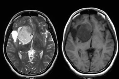
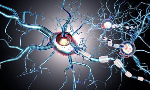
MS affects more than 1.8 million people worldwide and is characterised by lesions in the brain and spinal cord. In this condition, the body’s immune cells attack oligodendrocytes, which form myelin and belong to the group of glial cells. Signals between nerve cells cannot travel as quickly without myelin, which results in symptoms like reduced sensation and lack of coordination.
Co-first author and PhD student Petra Kukanja at the Karolinska Institutet said: “We wanted to understand which cells are part of the lesions and their dynamics over time.” Using in situ sequencing, developed in the research group of Professor Mats Nilsson at Stockholm University, the team could identify cells that are part of a tissue section by reading which genes are active in a particular cell. This pattern shows how different cell types are arranged in the tissue, in this case the spinal cord, as well as how the cells interact with each other.
Samples were taken from mice experimentally induced with MS-like symptoms and from human MS patients at multiple times to study how the lesions develop. Christoffer Mattsson Langseth is co-first author of the study and PhD student at the Department of Biochemistry and Biophysics, Stockholm University at Professor Mats Nilsson’s group. He explained: “We analysed simultaneously 239 genes and saw that active lesions in mouse were built up centrifugally in two dimensions, with immune cells in the middle and different types of glial cells around them.”
It was observed in mice that lesions first appeared in the spinal cord and then spread towards the brain. In samples of the spinal cord from four deceased MS patients, 260 genes were simultaneously analysed and the cellular architecture of the lesions could be determined. The authors found new lesions and new sub-structures within the lesions.
Kukanja said of oligodendrocytes: “The fact that they are active in the outer regions of lesions but also in the entire brain and spinal cord raises the question of whether they dampen the disease or drive it.”
In the future, the researchers plan to use the same methodology to analyse samples from more MS patients, as the disease is very heterogeneous. They also hope to investigate what the lesions look like when patients have received different types of treatments.
This study was published in Cell.