The mouse model provides a new understanding of the fundamental aspects of KSHV, which will enable drug and vaccine development.

Scientists at UNC Lineberger Comprehensive Cancer Center have developed a mouse model of Kaposi sarcoma after decades of research, that could be crucial for the development of new drugs to treat the disease.
Senior corresponding author Dr Dirk Dittmer, co-leader of the UNC Lineberger Virology Research Program and director of the UNC Viral Genomics Core, stated: “This is an important development as we have created the first animal model ever of Kaposi sarcoma. Animal models are essential to move new drugs from the laboratory bench into clinical trials.
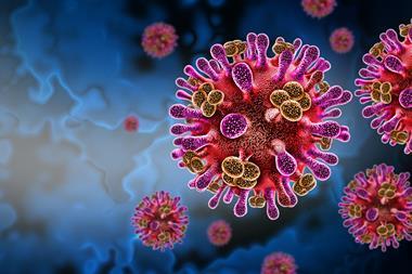
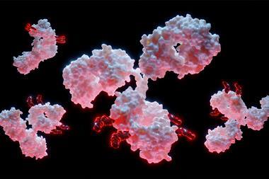
About 20 percent of all human cancers arise from viruses or require viral infection as an essential cofactor. Discovered in 1994, the Kaposi sarcoma-associated herpes virus (KSHV) is associated with Kaposi’s sarcoma as well as B-cell cancers. KSHV-associated diseases affect internal organs and are ultimately fatal. In the US, the diseases are present mainly in immunosuppressed people like those who are HIV-positive. An estimated 34,270 cases of Kaposi sarcoma were diagnosed globally, and 15,086 deaths were reported in 2020, with twice as many cases and deaths occurring in men compared to women. Africa accounted for 73 percent of new cases and 86.6 percent of the deaths from Kaposi sarcoma worldwide. In some southern and eastern African countries, the disease is endemic, and not HIV-related.
One method to study cancer is to observe tumour cells in the lab. However, Dr Dittmer said that Kaposi sarcoma tumour cells are very finicky and dependent on signalling molecules and blood supply, which is why they do not survive in a laboratory culture dish. This means that researchers have been required to develop animal models that would mimic, as closely as possible, the disease in humans.
A large challenge in developing the model was that two types of genes are transcribed into proteins in the Kaposi sarcoma mouse model. Normally when a virus infects a cell, the cell dies as the virus replicates, in a process named cell lysis. Cancer viruses are different as they enter a quiet state, called latency, where only the genes that help the infected cell survive are expressed. The mouse model the team developed is complex as both types of genes were needed.
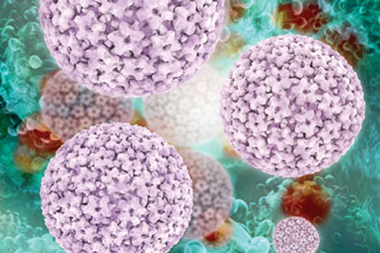
Cervical cancer and its related virus, HPV, offers a useful comparison for the challenge of developing a Kaposi sarcoma mouse model. The KSHV genome is 20 times larger than HPV. HPV has two cancer-causing genes, E6 and E7, so to mimic the disease in animals, researchers only needed to design two mice, one for each gene. As KSHV may have as many as 10 cancer-causing genes that all work together, it would be too challenging to develop that many mice.
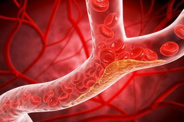
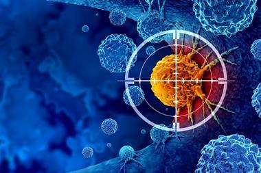
Dr Dittmer explained: “Another of the key virtues of our new mouse model is that it helps us understand angiogenesis, or new blood vessel formation. Without angiogenesis cancer cells are deprived of oxygen and die…In this mouse model, we can study angiogenesis blocking drugs better than ever before. If new drugs work against Kaposi sarcoma, they will also likely work against lesser-angiogenic tumours, which would be a major plus.”
The researchers hope that others will pursue drug and vaccine development based on a new understanding of fundamental aspects of KSHV provided by their mouse model. This includes the potential development of a primate model for human KSHV and Kaposi sarcoma.
This study was published in Cell Host & Microbe.