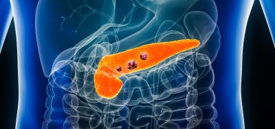
Contrast-enhanced subharmonic imaging detects prostate cancers
Posted: 16 April 2018 | Dr Zara Kassam (Drug Target Review) | No comments yet
A test of contrast-enhanced subharmonic imaging has shown promise in detecting prostate cancers that were not identified by MRI…
A test of contrast-enhanced subharmonic imaging (SHI) has shown promise in detecting prostate cancers that were not identified by MRI, according to a new study.
SHI is a new technique for imaging of microbubble ultrasound contrast agents with improved suppression of background tissues. The study carried out by Venkata Masarapu of Jefferson Medical College of Thomas Jefferson University was conducted to evaluate contrast-enhanced SHI of the prostate for detection of prostate cancer.
Among the study group of 28 patients, contrast enhancement was clearly observed with colour and power Doppler imaging, as well as with both harmonic imaging (HI) and SHI techniques in all subjects. SHI provided improved contrast signal and tissue suppression relative to conventional HI. Areas of increased vascularity were best delineated with maximum-intensity projection SHI, which allowed visualisation of microvascular architecture.
This first in vivo application of contrast-enhanced SHI demonstrated contrast enhancement in the prostate of every study patient, with focal areas of contrast enhancement corresponding to sites of cancer in 18% of targeted biopsies, including five patients whose cancer was not identified by MRI. The results indicate that SHI provides improved conspicuity of microbubble contrast enhancement and may improve the performance of contrast-enhanced ultrasound for detection of prostate cancer.
The research will be presented at the ARRS 2018 Annual Meeting, set for April 22-27 in Washington, DC.
Related topics
Imaging, Magnetic resonance images (MRI), Oncology
Related conditions
Prostate cancer
Related organisations
Jefferson Medical College
Related people
Venkata Masarapu