AI system detects cancer tumours missed by conventional diagnostics
Posted: 23 August 2018 | Iqra Farooq (Drug Target Review) | No comments yet
Computer engineers have ‘taught’ a computer to identify tiny specs of lung cancer in CT scan images, often missed by radiologists…
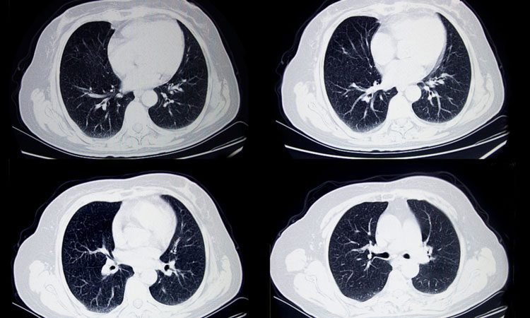
Engineers have used machine learning to ‘teach’ a computer how to detect tiny specs of lung cancer in CT scans.
Researchers from the University of Central Florida’s Computer Vision Research Centre taught the machine to identify areas often missed by radiologists, with an accuracy rate of 95 percent, compared to 65 percent by human eyes.
The researchers describe the method as being similar to algorithms used by facial-recognition software, where thousands of faces are scanned for patterns to identify the right match.
Biomarkers aren’t just supporting drug discovery – they’re driving it
FREE market report
From smarter trials to faster insights, this report unpacks the science, strategy and real-world impact behind the next generation of precision therapies.
What you’ll unlock:
- How biomarkers are guiding dose selection and early efficacy decisions in complex trials
- Why multi-omics, liquid biopsy and digital tools are redefining the discovery process
- What makes lab data regulatory-ready and why alignment matters from day one
Explore how biomarkers are shaping early drug development
Access the full report – it’s free!
Rodney LaLonde, a PhD student said: “We used the brain as a model to create our system. You know how connections between neurons in the brain strengthen during development and learn? We used that blueprint, if you will, to help our system understand how to look for patterns in the CT scans and teach itself how to find these tiny tumours.”
The National Institute of Health collaborated with Mayo Clinic and provided over 1,000 CT scan images, which the group fed into the software they developed. Post-graduate students worked on this study to ‘teach’ the computer different aspects to help it ‘learn’ what was needed to diagnose appropriately.
Assistant Professor Ulas Bagci lead the team, with PhD student Naji Khosravan creating the backbone of the whole system. Lalonde taught the computer how to ignore other tissue, nerves and masses encountered in the CT images, and to analyse lung tissues. Dr Sarfaraz Hussein fine-tuned the AI’s ability to identify cancerous and benign tumours.
“I believe this will have a very big impact,” Prof Bagci said. “Lung cancer is the number one cancer killer in the United States and if detected in late stages, the survival rate is only 17 percent. By finding ways to help identify earlier, I think we can help increase survival rates.”
The researchers are now looking to move the research into a hospital setting, and will present their research at the MICCAI 2018 conference in Spain, with their work published online.
Related topics
Artificial Intelligence, Disease Research, Oncology, Research & Development, Screening
Related conditions
Lung cancer
Related organisations
Computer Vision Research Centre, University of Central Florida
Related people
Assistant Professor Ulas Bagci, Dr Sarfaraz Hussein, Naji Khosravan, Rodney LaLonde