Innovative mechanobiology research expands understanding of cells
Posted: 13 May 2019 | Drug Target Review | No comments yet
Researchers have developed a new technology that allows them to probe cell changes without disturbing the cell’s physiology.
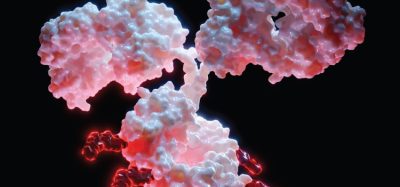
This is a major advancement that can aid scientists in looking more closely at cell changes to solve human health problems, according to a new paper in Cell Reports by researchers from the University of Colorado Boulder.
The paper was written by CSU Professor Soham Ghosh as first author, CU Professor Corey P Neu, CU graduate student Benjamin Seelbinder and other contributors. “This technology can drive the field of mechanobiology at an unprecedented rate,” said Professor Ghosh. “The technique has proven powerful in several applications and is opening new avenues of research.”
This technology, known as Deformation Microscopy, allows scientists to more accurately assess the interplay between biological systems and the mechanical environment in treating musculoskeletal, cardiac and neural diseases.
Advancements in regenerative medicine and developmental-biology-inspired technology have the potential to target major human health obstacles. With the team’s discovery, scientists can get a clearer understanding of how the physical changes in cells contribute to cell development – an area of study known as mechanobiology.
Before joining CSU 2018, Ghosh and his colleagues developed the technology to non-invasively probe the mechanics of the biological system in high resolution using an image-based formula. The results showed unhindered images of the cell, and inside the cell nucleus, exposing the missing link between biological systems and the mechanical environment. By viewing and analysing the cell and nucleus at high magnification, all the intricate structural architectures became visible.
This helped researchers understand spatially detailed and dynamic cell deformation, which subsequently revealed how the subcellular and subnuclear regions behave in a normal physiological setting versus a diseased setting.
Mechanical probing in biology has been prevalent for almost two decades. Previous technologies were invasive, disturbing the physiology of the cell. Deformation Microscopy produces a high-precision, microscopic image-based strain map which can quantify the affliction a cell and nucleus can sustain, revolutionising what was previously known about mechanobiology.
Ghosh will leverage a variation of Deformation Microscopy and other methods to quantify biological phenomena in cells in his Cellular Engineering and Mechanobiology Lab, to investigate questions surrounding single cell changes in relation to stem cell function. Researchers can investigate, for example, why individual tissues are functionally different despite having the same genetic code.
His lab will study the interaction between the cell’s immediate environment, epigenetics, and chromatin architectural organisation of the cell nucleus.
Related topics
Analytical Techniques, Cell Cultures, Imaging, Technology
Related organisations
University of Colorado Boulder.
Related people
Benjamin Seelbinder, Corey P Neu, Professor Soham Ghosh