Free AI platform created to analyse microscopy images
Posted: 26 April 2021 | Victoria Rees (Drug Target Review) | No comments yet
An AI software called ZeroCostDL4Mic has been developed by researchers to enable other scientists to analyse images from microscopy studies.
Researchers have developed a new artificial intelligence (AI) software that can be used in the processing of bioimaging data. The technology was created at Åbo Akademi University, Finland.
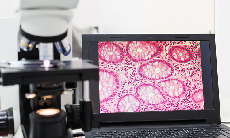
The team say that software using AI is revolutionising how microscopy images are analysed. For instance, AI can be used to detect features in images (ie, tumours in biopsy samples) or improve the quality of images by removing unwanted noise. The researchers describe a platform called ZeroCostDL4Mic, which can make these AI technologies accessible to everyone.
“The key novelty is that ZeroCostDL4Mic runs in the cloud for free and does not require users to have any coding experience or advanced computational skills. Effectively, it runs on any computer that has a web browser,” said Guillaume Jacquemet, one of the researchers from the study.
The team highlight that microscopy is a leading technology used worldwide to perform not only research but also diagnostics.
Modern microscopes are directly connected to digital cameras, leading to the acquisition of hundreds to thousands of images per sample. These images need to be processed on a computer to gain meaningful data, which is a huge undertaking. However, non-experts can continue to find AI technologies difficult to use.
To help with the number of images, the researchers used AI to train a machine to do the work. In practice, ZeroCostDL4Mic is a collection of self-explanatory notebooks for Google Colab, featuring an easy-to-use graphical user interface.
“We believe that ZeroCostDL4Mic will acts as ‘a gateway drug’ for AI, luring users to explore these new technologies that will transform biomedical research and diagnostics in the decades to come,” said Jacquemet.
The paper from the study is published in Nature Communications.
Related topics
Artificial Intelligence, Imaging, Informatics, Lab Automation, Microscopy
Related organisations
Åbo Akademi University
Related people
Guillaume Jacquemet