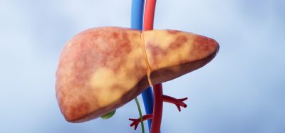
Scientists create advanced 3D liver model mimicking real tissue
Posted: 5 June 2025 | Drug Target Review | No comments yet
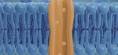
Scientists have developed a 3D liver model, known as the periportal assembloid. This model replicates the liver’s complex structure and bile transport system, enabling more precise study of disease progression.

In a new study published in Nature, researchers from the Max Planck Institute of Molecular Cell Biology and Genetics (MPI-CBG) have developed a sophisticated 3D liver model called the periportal assembloid. This model accurately reconstructs the liver’s periportal region, incorporating the three main cell types essential for liver function- hepatocytes, cholangiocytes, and mesenchymal cells. The assembloid mimics the liver’s architecture and bile transport system with direct precision, enabling scientists to study bile flow, cholestatic injury, and fibrosis in a lab setting. This innovative model marks a major step forward in understanding liver disease mechanisms and testing drug responses in a more physiologically relevant environment.
Laying the groundwork: previous advances in liver organoids
In 2021, a team led by Dr Meritxell Huch, director at the Max Planck Institute of Molecular Cell Biology and Genetics (MPI-CBG), made significant progress toward addressing these limitations. Their earlier work produced a dual-cell liver organoid composed of cholangiocytes (bile duct cells) and mesenchymal cells. This model succeeded in replicating some cell-to-cell interactions but still lacked hepatocytes- the central functional cells of the liver that make up most of its mass.
Creating a next-generation liver model: the periportal assembloid
This new model includes hepatocytes, cholangiocytes, and mesenchymal cells, making it the first lab-grown system to mimic the liver’s periportal region.
Biomarkers aren’t just supporting drug discovery – they’re driving it
FREE market report
From smarter trials to faster insights, this report unpacks the science, strategy and real-world impact behind the next generation of precision therapies.
What you’ll unlock:
- How biomarkers are guiding dose selection and early efficacy decisions in complex trials
- Why multi-omics, liquid biopsy and digital tools are redefining the discovery process
- What makes lab data regulatory-ready and why alignment matters from day one
Explore how biomarkers are shaping early drug development
Access the full report – it’s free!
“Our assembloid reconstructs the liver periportal region and can model aspects of cholestatic liver injury and biliary fibrosis,” says Anna Dowbaj, co-first author and soon-to-be assistant professor at the Technical University of Munich (TUM). “We chose this region in particular since it plays a key role in bile transport and is often disrupted in liver diseases when the connection of cells responsible for bile transport is blocked.”
Functionality and disease modelling in the lab
The researchers first developed organoids composed exclusively of hepatocytes, which formed functional bile channels. These hepatocytes retained key features of liver tissue. By then adding cholangiocytes and fibroblasts, they were able to build full periportal assembloids.
“Our liver model works like real liver tissue, moving bile from inside the liver cells into bile ducts,” explains Aleksandra Sljukic, doctoral student and co-first author. “This shows that we were able to replicate the interactions between the different liver cells.”
Our liver model works like real liver tissue, moving bile from inside the liver cells into bile ducts.
Importantly, by adjusting the number of mesenchymal cells, researchers could simulate fibrosis-like responses. They also used genetic manipulation techniques, such as mixing normal and mutant cells or deactivating specific genes, to study liver disease progression.
Additionally, using topological data analysis, Harrington’s team at the University of Oxford was able to categorise assembloid shapes. Certain shapes were associated with better liver function over time, offering new ways to assess organoid health and development.
A promising tool for future research and drug development
Huch highlights the importance of this new model:
“We are excited that we were able to create a periportal assembloid model that combines, for the first time, portal mesenchyme, cholangiocytes, and hepatocytes. Although some cells are still missing, namely the endothelium and immune cells, the model captures with high precision the cellular and structural architecture of the liver’s periportal area at the scale of a tissue culture dish.”
She notes that its modular design makes the model easily adaptable and accessible for lab research.
“Our liver assembloids are the first all-in-one lab model capable of studying bile flow, bile duct injury, and how different liver cells contribute to disease.”
Looking ahead, the team envisions even broader applications for the future.
“Once translated to human cells, it could be a way of moving from 2D models utilised in pharmaceutical screenings to more physiological 3D models to study drug efficacy and toxicity in a more physiologically relevant context.”
Conclusion
This study represents a major advance in liver disease modelling and regenerative medicine. The periportal assembloid deepens our understanding of liver function and disease progression. It could also support the development of more effective personalised treatments and drug testing platforms. As research continues, the inclusion of additional cell types will further enhance the model’s relevance and potential in biomedical science.
Related topics
Cell Cultures, Cell Line Development, Disease Research, Drug Discovery, Drug Discovery Processes, Hepatocytes, Organoids, Regenerative Medicine
Related conditions
Liver disease
Related organisations
Max Planck Institute of Molecular Cell Biology and Genetics (MPI-CBG)
Related people
Aleksandra Sljukic (Doctoral Student at Max Planck Institute of Molecular Cell Biology and Genetics (MPI-CBG)), Anna Dowbaj (Assistant professor at the Technical University of Munich (TUM)), Dr Meritxell Huch (Director at the Max Planck Institute of Molecular Cell Biology and Genetics (MPI-CBG))