
Featured content

Article
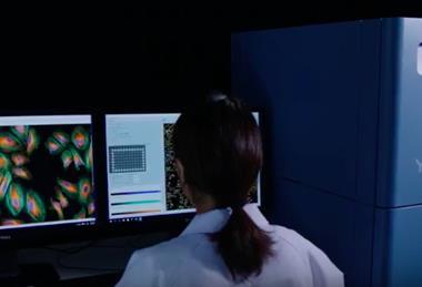
Operational data: the hidden driver of faster drug discovery
The key to faster, smarter drug discovery lies in data that’s often overlooked. By exposing hidden delays and inefficiencies, this data enables teams to shorten discovery cycles and progress promising candidates faster.
 Article
From fragments to maps: scaling drug–target interaction data
Most drug–target data were never designed to be compared at scale. Pharmome mapping takes a different approach, building a shared dataset intended to support more predictable discovery.
 Interview
Computational design drives new generation of synthetic promoters
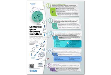
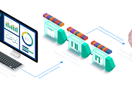
Designing gene control from scratch is becoming possible. SynGenSys is using computational design to create synthetic promoters for advanced therapies.
 Interview
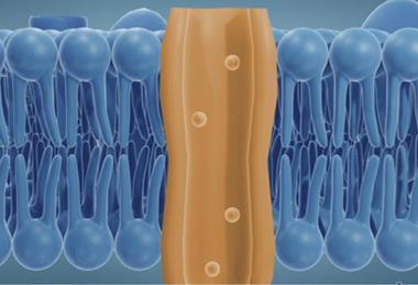
Physics-based modelling offers a new way to study drug targets
Australian start-up OmnigeniQ has demonstrated what it describes as the first deterministic, physics-based computation of a human protein in its native state.
Opinion and interviews
Computational design drives new generation of synthetic promoters
Designing gene control from scratch is becoming possible. SynGenSys is using computational design to create synthetic promoters for advanced therapies.
Opinion and interviews
- Previous
- Next


Join now
Unlock exclusive industry insights
- Bookmark articles and resources to access anytime
- Enjoy free access to industry leading resources, webinars and insights
- Stay informed with the latest news and breakthroughs
- Receive updates and recommendations tailored to your research interests
Webinars
- Previous
- Next