New method for developing medical imaging tracers discovered
Posted: 21 June 2019 | Drug Target Review | No comments yet
A new medical imaging tracing method has been discovered by scientists which can better track drugs in the body.
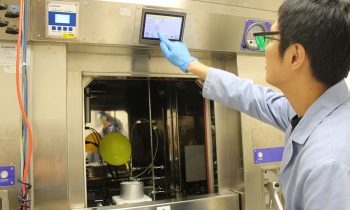
Scientists from the University of North Carolina Lineberger Comprehensive Cancer Centre have discovered a method for creating radioactive tracers to better track pharmaceuticals in the body as well as image diseases and other medical conditions.
The method is for creating tracers which can be used with positron emission tomography (PET) imaging, making it possible to attach radioactive tags to compounds that previously have been difficult or even impossible to label.
“Positron emission tomography is a powerful and rapidly developing technology that plays key roles in medical imaging as well as in drug discovery and development,” said the study’s co-corresponding author, UNC Lineberger’s Zibo Li, PhD, an associate professor in the UNC School of Medicine Department of Radiology, and director of the Cyclotron and Radiochemistry Program at the UNC Biomedical Research Imaging Centre. “This discovery opens a new window for generating novel PET agents from existing drugs.”
The researchers have described a new way of attaching the radioactive molecule Fluorine-18 (a widely used isotope in PET imaging) by breaking a specific chemical structure of carbon and hydrogen atoms in their study. In the presence of blue light from a laser and after the addition of a catalyst material to speed the reaction, the researchers could break existing chemical bonds in the structure and insert Fluorine-18. Once attached, the tracer emits gamma rays that are picked up by imaging.
They envision multiple potential applications for their discovery, including for medical imaging to screen patients for response to a drug, or to aid in drug development research. They have also stated their approach may allow them to attach a tag to existing compounds: “In this study, we showed that we could label a broad spectrum of compounds,” Li continued, including for anti-inflammatory drugs, and specific amino acids to show that they could image tumours.
The researchers said their next step is to develop a device that would make it easier for scientists to use this new method for creating radiolabeled tracers. In addition, they are working to expand their technology to develop other tracers that use a different radioactive material, such as Carbon-11.
The researchers have reported their method in the journal Science.
Related topics
Amino Acids, Drug Development, Imaging, Research & Development
Related organisations
North Carolina Lineberger Comprehensive Cancer Centre
Related people
Zibo Li PhD