Spatial biology is the next life science revolution
Posted: 17 June 2022 | Dr Richard Goodwin (AstraZeneca) | No comments yet
Dr Richard Goodwin, Head of Imaging & AI, Clinical Pharmacology & Safety Sciences at AstraZeneca, explores the latest innovation in early therapeutic development – spatial biology.
Those researching life sciences will recognise that technological invention drives innovation. This can be through game changing techniques like CRISPR gene editing, where seismic shifts in capabilities can open up a new research landscape. Innovation can also be the culmination of new technologies alongside the refinement of established techniques, which through advancements in artificial intelligence (AI) allow us to explore complex data, driving progress forward. Such innovation “evolution” can occur over a number of years and then be supercharged by the final piece of the jigsaw puzzle slotting into place – then we have a revolution. We are seeing this today with the spatial biology revolution. It is changing how we think about our patients, their disease heterogeneity within the wider population, context between immune response, disease progression, therapeutics response and their interplay across the tissue microenvironment.
You may also like:
Drug discovery from the age of information to the age of intelligence
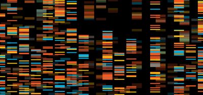
Spatial biology can be defined many ways, but simply put it is the study of the tissue microenvironment in unprecedented detail. Moving beyond a morphological assessment of the tissue to one that incorporates extensive quantitative evaluation of cell phenotypes and biomarker expression to understand how cells interact and respond within disease progression or on treatment. It moves away from “hypothesis targeted” measurement of a sample, to one that generates vast data multiparametric assessment that also considers biomarker contextualisation and localisation.
Spatial biology is the logical evolution of bulk to single-cell analysis to whole-tissue analysis. Moving beyond the homogenate measurements – still so informative in providing high-throughput averaged protein, metabolite or genomic information – to those able to analyse single cells and reveal their unique phenotypes. Then, ultimately, making similar biomarker measurements but spatially resolved (Nature Methods method of the year: next-generation sequencing in 2009, mass spectrometry‑targeted proteomics in 2012, single cell genomics in 2013, single cell multimodal omics in 2019, spatially resolved transcriptomics in 2020).
Researchers should not be limited by what can be measured using a single technology platform. Rather, they should be able to explore how different spatially resolved biomarker technologies are compatible on the same formalin-fixed or fresh frozen tissue sample. Knowing the hidden heterogeneity within patient biopsies, then disease models, is the only way to ensure we are designing the right medicines for the right patient population. Does this mean we can expect to see deep tissue phenotyping on every patient sample of pre-clinical study? No, the idea is to perform deep exploration using as many compatible technologies as possible, integrating the data and then extracting biomarkers that informs sufficiently, but at higher capacity. This is reflected by the Rosetta stone analogy exemplified by Cancer Research UK Grand Challenge team.1 Metabolic mass spectrometry imaging measurements are incorporated with spatial transcriptomics, multiplex immunohistochemistry (IHC) for cell phenotyping and connected with advanced digital pathological view of the same tissue.
What does this deliver to innovative companies, like AstraZeneca, that embed spatial biology into all parts for the R&D pipeline? Opportunities to use AI data-driven routes to segmentation beyond genetics and bulk RNA profiling, understanding heterogeneity to improve outcome predictions, novel routes to personalised medicine and diagnostics and ultimately improved platform of evidence (TI to clinical concept testing).
About the author
Dr Richard Goodwin is head of Imaging & AI, Clinical Pharmacology & Safety Sciences at AstraZeneca. He leads a global team of imaging scientists that deliver expertise across the company using cutting‑edge multidimensional imaging modalities. Richard derives project insight and impact by connecting safety and efficacy endpoints using advanced tissue histology, molecular imaging, in vivo PET, MRI and much more. He has published over 60 publications while at AstraZeneca on the development and deployment of new technology in support of the portfolio. Richard is also an Honorary Professor at the University of Glasgow.
Reference
- Rosetta | Cancer Grand Challenges [Internet]. Cancergrandchallenges.org. 2022 [cited 30 May 2022]. Available from: https://cancergrandchallenges.org/teams/rosetta
Related topics
Analysis, Bioinformatics, Biomarkers, Informatics, Molecular Biology, Personalised Medicine, Technology
Related organisations
AstraZeneca, Cancer Research UK (CRUK)