A growing body of evidence pinpoints neuroinflammation as a pivotal factor driving brain-related pathogenesis. Yet, a crucial question lingers: among the various immune cell groups residing within the brain, which one orchestrates this inflammatory reaction?
A recently published study in Nature Communications led by Dr Ashley Harms and her team at the University of Alabama at Birmingham explores further. By employing a mouse model of Parkinson's disease, the researchers have illuminated a surprising revelation: it's the border-associated macrophages (BAMs), not the microglia, that steer the neuroinflammatory response within the brain. This discovery bears significance since prior research has already shown that neuroinflammation exacerbates neurodegeneration in this particular Parkinson's mouse model.
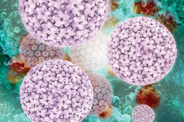
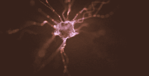
In humans, the abnormal build up of the protein alpha-synuclein culminates in the demise of brain neurons responsible for producing the neurotransmitter dopamine. In the mouse model, where human alpha-synuclein is overly produced in dopaminergic neurons, many of the inflammatory characteristics witnessed in human disease are replicated.
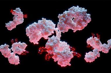
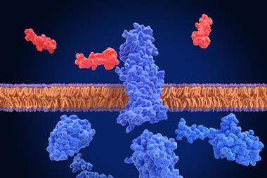
Two distinct subsets of macrophage immune cells stationed within the brain are microglia and border-associated macrophages (BAMs), also known as central nervous system-resident macrophages (CRMs). These macrophages, a kind of white blood cell, engulf and digest foreign intruders like cancer cells and bacteria. Following this, they play a vital role in stimulating the adaptive immune response for more robust defence by displaying fragments of protein from the intruders, known as antigens, on their surface. These antigens, showcased on macrophages by a carrier protein called MHCII, activate T cells, pivotal components of the immune response. Prior research had indicated the pivotal role of MHCII in the inflammation and degeneration associated with Parkinson's disease. However, identifying the precise cells presenting the antigens remained elusive.
The UAB team showed that removing MHCII from CRMs, encompassing both microglia and BAMs, safeguarded against the loss of dopaminergic cells in the mouse model. This suggested that antigen presentation specifically by CRMs is fundamental for the inflammation and ensuing neurodegeneration driven by alpha-synuclein.
Interestingly, deleting MHCII in microglia alone did not diminish neuroinflammation, leading the researchers to deduce that some other non-microglial subset of CRMs must be responsible for presenting the alpha-synuclein antigen to CD4+ T cells, thus prompting neuroinflammation and the influx of peripheral immune cells from outside the central nervous system into the brain.
In contrast, selectively reducing BAMs specifically resulted in decreased indicators of neuroinflammation. Harms discusses how these findings indicate that BAMs, not microglia, are required for peripheral immune cell recruitment into the brain parenchyma and antigen re-stimulation, a process responsible for alpha-synuclein-induced neurodegeneration.
The researchers also observed that heightened alpha-synuclein expression led CRMs to transform into disease-associated microglia and disease-activated BAMs. This transformation was characterised by specific patterns of gene activation related to functions such as cytokine signalling and phagocytosis. This activation resembled that of previously identified activated CRMs in conditions like Alzheimer's disease and amyotrophic lateral sclerosis.
Additionally, the study aimed to bridge the gap between the mouse findings and human Parkinson's disease. By comparing post-mortem mid-brain samples of Parkinson's patients with control samples, the researchers found that Parkinson's patients exhibited a significantly higher proportion of T cells, including CD4+ and CD8+ T cells, adjacent to BAMs compared to healthy controls. Harms explains how this mirrors what we see in our mouse model of Parkinson's disease, indicating a disease-associated interaction similar to that observed in mice.
This insight shifts our conventional understanding of neuroinflammatory mechanisms in neurodegenerative diseases. It underscores BAMs' unique and non-redundant functions in terms of immune cell recruitment, class II antigen presentation, and T cell infiltration. According to Harms, these results underscore BAMs' critical role in neuroinflammatory responses, emphasising the necessity for therapeutic targeting of BAMs to modify disease progression in neurodegenerative disorders. Notably, BAMs are so-named because of their proximity to the blood vessels at the borders of the central nervous system.