Distinct macrophage signatures that align specifically with regeneration could offer novel therapies to enhance wound healing.
Scientists from Cincinnati Children’s Hospital and the University of Kentucky have investigated how spiny mice can regenerate lost tissue. They are using this new understanding to initiate regeneration in other types of mice, which could be translated into humans in the future.
Adult laboratory mice heal injuries with scar tissue, but spiny mice have the unique ability to regrow lost skin and regenerate musculoskeletal tissues in their body. Dr Ashley Seifert, an associate professor in the Department of Biology at the UK College of Arts and Sciences, and his research group have pioneered the use of spiny mice and other animal models to understand how complex tissue can regenerate, bridging regenerative biology and medicine.
A study1 from Seifert’s group, published last year, demonstrated how ERK signalling acts as a crucial switch balancing the healing response. It also showed that increasing and maintaining this type of signalling in laboratory mice could initiate a regenerative response instead of scarring.
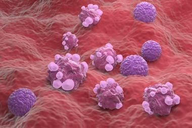
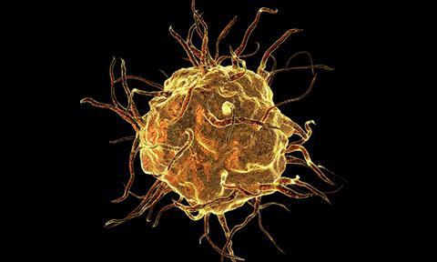
Macrophages
In the new study, the researchers offer novel insights into how specific immune cells respond to injury and direct tissue regeneration. They studied macrophages, part of the innate immune system which recognise, engulf and destroy many potential pathogens. Their destructive potential and ability to secrete regulators of the function of neighbouring cells contribute to many aspects of homeostasis.2 They also promote tissue repair in both spiny mice and lab mice.
Dr Seifert explained: “In our study, we used a dual-species system to dissect macrophage phenotypes between two modes of healing: tissue regeneration or scar tissue formation…While it’s still unclear the extent to which macrophages ultimately control different healing trajectories, we observed subtle and distinct macrophage signatures that aligned specifically with regeneration.”
“Overall, our experiments point towards macrophages establishing a tissue microenvironment conducive for regeneration.”
The team observed the signals released by macrophages that communicate to nearby cells, using an identical injury in both types of mice. They noted the phenotypes of the cells during those processes. “Our study shows that spiny mouse macrophages release distinct proteins that are partially responsible for the reformation of specialised tissues at the site of injury and for protecting cells from stress,” revealed Dr Seifert. “Overall, our experiments point towards macrophages establishing a tissue microenvironment conducive for regeneration.”
The macrophages from the bone marrow of both types of mice were examined and compared. Using RNA sequencing to identify the proteins these cells secrete, they discovered that the macrophages from bone marrow responded differently on their own to interferon gamma, a protein produced by cells in response to infection, and lipopolysaccharide, a molecule found in some bacteria that stimulates the immune system.
Regenerative healing
Dr Seifert elucidated: “In response to inflammatory cues, macrophages in spiny mice release unique communicating proteins that promote the growth of new blood vessels, lymphatic vessels, anti-inflammatory activities and tissue rebuilding that positively contribute to regenerative healing.”
The UK team found that those cells maintain their uniqueness throughout scar tissue formation or regeneration. Samples at five different time points during healing were studied to better understand the timeline for how macrophages carry out their work. A gene analysis showed researchers which types of molecules produced by the cells were most active in each type of mouse and with which functions those were most associated.
Dr Seifert commented: “In our work to answer more scientific questions about spiny mice, we found that a specific type of protein — vascular endothelial growth factor c or VEGFC — is uniquely secreted by spiny mouse macrophages during regeneration…It also plays a multifunctional role, encouraging the growth of new blood and lymphatic vessels.”
“Through specific antibody blocking of VEGFC protein in the ears of spiny mice, we observed notable changes in the formation of blood vessels, lymph vessels and cell division."
Co-first author of the study and a postdoctoral scholar in biology Dr Ajoy Aloysius explained: “Through specific antibody blocking of VEGFC protein in the ears of spiny mice, we observed notable changes in the formation of blood vessels, lymph vessels and cell division. This also led to decreases in new hair follicle formation and increases in inflammation, ultimately disrupting the process of tissue regeneration.”
He added: “Additionally, we speculate that enhancing the secretion of VEGFC and other factors by macrophages during fibrotic wound healing may contribute to a regenerative outcome in tissue healing, however further experiments are required to confirm this idea.”
Future applications
The results suggest that macrophages in specific tissues throughout the body aid the direction and regulation of the cellular repair programme. The type of macrophage is significant, as altering the secretion of one type of macrophage could change how tissue repairs itself.
One of the study’s first authors Dr Jennifer Simkin, an assistant professor of orthopaedic surgery at the Louisiana State University Health Sciences Center, concluded: “This study aims to unlock the body’s natural potential to regenerate after traumatic injury…Ultimately, we hope these findings pave the way for novel therapies to enhance wound healing.”
Further study is required for an improved understanding and definition of some of this cellular crosstalk during the regeneration process.
This new study was published in Developmental Cell.
References
1 Bartscherer K, Koopmans T, Linjnzaad P. An ERK-dependent molecular switch antagonizes fibrosis and promotes regeneration in spiny mice (Acomys). 2023 April 26 [2024 January 26]; 9(17). Available from: https://www.science.org/doi/10.1126/sciadv.adf2331
2 Hume DA, Sasmono RT. The Innate Immune Response to Infection [Internet]. Chapter 4: The Biology of Macrophages. Wiley Online Library. 22 June 2004 [2024 January 26]. Available from: https://doi.org/10.1128/9781555817671.ch4