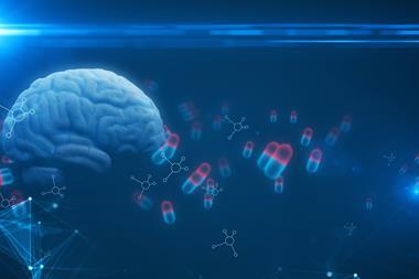
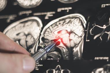
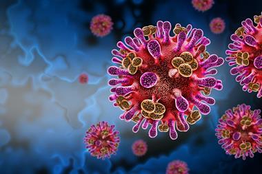
A form of MRI is a better alternative to many existing imaging technologies when looking at how COVID-19 can change the human brain.
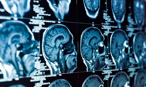
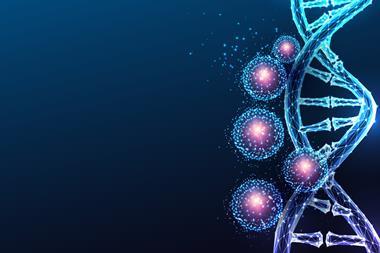
Systems Design Engineering Professor Alexander Wong, who is also the Canada Research Chair in Artificial Intelligence and Medical Imaging, had previously developed correlated diffusion imaging (CDI) in a successful search for a better imaging measure for detecting cancer.
CDI is a new form of MRI that can better highlight the differences in the way water molecules move in tissue by capturing and mixing MRI signals at different gradient pulse strengths and timings. The imaging technique out of the University of Waterloo, UK, has recently been used in a new study, published in Human Brain Mapping, by scientists at Baycrest’s Rotman Research Institute and Sunnybrook Hospital in Toronto, Canada.
“Some may think COVID-19 affects just the lungs,” Wong explained. “What was found is that this new MRI technique that we created is very good at identifying changes to the brain due to COVID-19. COVID-19 changes the white matter in the brain.”
Researchers at Rotman, a world-renowned centre for the study of brain function, saw Wong’s imaging discovery and thought it could likely also be used to identify changes to the brain due to COVID-19.
Subsequent tests proved that theory right.
CDI imaging of frontal-lobe white matter revealed a less restricted diffusion of water molecules in COVID-19 patients. At the same time, it showed a more restricted diffusion of water molecules in the cerebellum of patients with COVID-19.
Wong highlights that the two regions of the brain react differently to COVID-19 and points to two key findings from the research. First, the human cerebellum might be more vulnerable to COVID-19 infections. Second, the study reinforces the idea that COVID-19 infections can lead to changes in the brain.
Not only is the study one of the few to have shown COVID-19’s effects on the brain, but it is the first to report diffusion abnormalities in the white matter of the cerebellum. Although the study was designed to show changes, rather than specific damage, to the brain from COVID-19, its final report does discuss potential sources of such changes and many link to disease and damage.
In response, Wong suggests future tests could focus on whether COVID-19 actually damages brain tissue. Additional studies could also determine if COVID-19 can change the brain’s grey matter.
“Hopefully, this research can lead to better diagnoses and treatments for COVID-19 patients,” Wong concluded. “And that could just be the beginning for CDI as it might be used to understand degenerative processes in other diseases such as Alzheimer’s or to detect breast or prostate cancers.”