Low-dose positron emission mammography (PEM) has a high sensitivity for detecting cancer and reduces the likelihood of false positives.

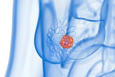
Researchers from the Radiological Society of North America (RSNA) have developed a breast imaging technique which provides high sensitivity for detecting cancer while greatly reducing the likelihood of false positive results. This promises more reliable breast cancer screening for a wider range of patients.
Although mammography is an effective screening tool for early detection of breast cancer, its sensitivity is reduced in dense breast tissue, because of the masking effect of overlying dense fibroglandular tissue. Almost half of the screening population has dense breasts, so many of these patients need additional breast imaging, often with MRI, after mammography.
Low-dose positron emission mammography (PEM) is a new molecular imaging technique that gives an improved diagnostic performance at a radiation dose comparable to that of mammography.
In the study, 25 women with a median age of 52 years, recently diagnosed with breast cancer, underwent low-dose PEM with the radiotracer fluorine 18-labeled fluorodeoxyglucose (18F-FDG). The PEM images taken one and four hours post 18F-FDG injection were reviewed by breast radiologists. These findings were correlated with lab results.
PEM displayed comparable performance to MRI, identifying 24 of the 25 invasive cancers. Notably, its false positive rate was only 16 percent, compared with 62 percent for MRI. The study also showed that PEM could decrease downstream healthcare because it may prevent further unnecessary work up compared to MRI. Also, the technology does not require breast compression, which is often uncomfortable for patients, as it is designed to deliver a radiation dose comparable to that of traditional mammography without it.
Lead author Dr Vivianne Freitas is assistant professor at the University of Toronto, and staff radiologist of the Breast Imaging Division of the Toronto Joint Department of Medical Imaging, University Health Network, Sinai Health System and Women’s College Hospital. She explained: “The integration of these features—high sensitivity, lower false-positive rates, cost-efficiency, acceptable radiation levels without compression, and independence from breast density—positions this emerging imaging modality as a potential groundbreaking advancement in the early detection of breast cancer.”
She continued: “As such, it holds the promise of transforming breast cancer diagnostics and screening in the near future, complementing or even improving current imaging methods, marking a significant step forward in breast cancer care.”
According to Dr Freitas, low-dose PEM offers clinical uses in both screening and diagnostic settings. “For screening, its ability to perform effectively regardless of breast density potentially addresses a significant shortcoming of mammography, particularly in detecting cancers in dense breasts where lesions may be obscured…It also presents a viable option for patients at high risk who are claustrophobic or have contraindications for MRI.”
PEM could also have a significant role in interpreting uncertain mammogram results, evaluating the response to chemotherapy and deciding the extent of disease in newly diagnosed breast cancer, including involvement of the other breast.
Dr Freitas believes that if PEM successfully lowers these rates, it could significantly lessen the emotional distress and anxiety linked to false positives, as well as decreasing unnecessary biopsies and treatments.
More studies are needed to determine low-dose PEM’s exact role and efficacy in the clinical setting. “While the full integration of this imaging method into clinical practice is yet to be confirmed, the preliminary findings of this research are promising, particularly in demonstrating the capability of detecting invasive breast cancer with low doses of fluorine-18-labeled FDG,” Dr Freitas concluded. “This marks a critical first step in its potential future implementation in clinical practice.”
This study was published in Radiology: Imaging Cancer.