
UCLA scientists have created the first detailed map of how the ovarian reserve forms in primates, offering new insights – and potential new treatments – for infertility, polycystic ovary syndrome (PCOS) and hormone-related conditions.
UCLA scientists have developed the first comprehensive road map showing how the ovarian reserve forms in primates, providing new insights into women’s health that could lead to the development of new treatments for infertility and polycystic ovary syndrome (PCOS).
The research, published in Nature Communications, is a six-year collaboration among scientists from UCLA, Harvard, UC San Francisco and the National Institutes of Health-funded Oregon National Primate Research Center.
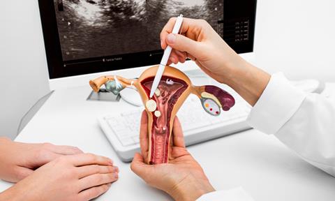
The ovarian reserve – the lifetime supply of eggs that a woman is born with – serves not only as the foundation for reproduction but also as the driver of hormone production in the ovaries.
“It’s what enables women to become mothers, girls to progress through puberty and acts like a biological clock counting down to menopause,” said senior author Amander Clark, a professor of molecular, cell and developmental biology and a member of the Eli and Edythe Broad Center of Regenerative Medicine and Stem Cell Research at UCLA. “We now have a manual that could help scientists create more accurate human ovarian models to better study ovarian disease and dysfunction.”
A model that mirrors human ovarian reserve development
Despite governing so many crucial aspects of women’s health, how this limited supply of eggs actually develops has remained a mystery.
The biggest obstacle has been access. In humans, the ovarian reserve forms entirely before birth – which is an extremely difficult window of time to study.
In humans, the ovarian reserve forms entirely before birth – which is an extremely difficult window of time to study.
To overcome this obstacle, the research team turned to the rhesus macaque, a primate that shares about 93 percent of its DNA with humans and undergoes remarkably similar ovarian and ovarian reserve development.
“We needed a model that has similar physiology to humans,” said first author Sissy Wamaitha, a postdoctoral scholar in the Clark lab. “And we know from historical studies that the various steps of ovarian reserve formation in primates are very similar to what occurs in humans.”
The researchers identified critical stages in ovarian reserve development, including initial ovary formation, female sex determination and follicle formation – the process by which protective sacs develop around eggs within the ovaries to support their survival.
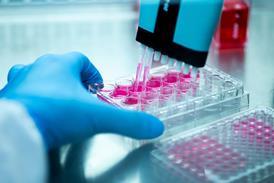
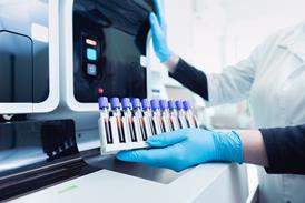
Using single cell sequencing and spatial transcriptomics technologies, they then analysed these developmental snapshots at the cellular and molecular level, starting from some of the earliest stages of ovarian reserve development.
“Women’s health is already understudied, but the ovary in particular has been neglected,” Wamaitha said. “To effectively treat reproductive health conditions – as well as the growing number of general health issues we now acknowledge affect people with ovaries – we must first develop a fundamental understanding of the full scope of this organ’s function.”
Solving the mini-puberty mystery
One of the study’s key findings provides the first cellular explanation for mini-puberty – a mysterious hormone surge that occurs in babies soon after birth.
Scientists observed that specialised hormone-producing cells activate in the ovary shortly before birth and this period of what Wamaitha calls 'practice growth' is responsible for the hormone spike detected during mini-puberty.
For infants who don’t experience mini-puberty, the absence of this hormone surge could serve as an early biomarker for ovarian dysfunction, such as PCOS – which affects approximately 10 percent of women worldwide.
“If we can identify risk factors in infancy that impact ovarian health, then early interventions can be made so that these women don’t suffer once they go through puberty,” said Clark.
Next steps: building better ovarian models
The first-of-its-kind atlas has immediate applications for stem cell researchers who have wanted to grow more accurate ovarian organoids in the lab.
In the past, these efforts were hindered because scientists lacked the detailed information needed to confirm they were creating the correct specialised cell types needed for ovarian reserve formation.
With this new strategy, the research team is already working to create the essential ovarian support cells from induced pluripotent stem cells.
If successful, they can combine these engineered support cells with lab-grown germ cells to create sophisticated 3D ovarian models – developing our understanding of infertility causes and accelerating the development of new treatments.
“This project demonstrates the value of basic research,” Clark said. “We’re advancing knowledge about an understudied organ to create tools that could meaningfully improve the health of women and girls everywhere.”


Topics
- Amander Clark (Professor Member of the Eli and Edythe Broad Center of Regenerative Medicine and Stem Cell Research at UCLA)
- Analytical Techniques
- Biomarkers
- Cell & Gene Therapy
- Disease Research
- Drug Discovery Processes
- Endocrine disorders
- Gene Therapy
- Genomics & Sequencing
- Harvard
- Industry Experts
- Molecular Biology
- Organoids
- polycystic ovary syndrome (PCOS)
- Regenerative Medicine
- Sequencing
- Sissy Wamaitha (Postdoctoral Scholar in the Clark Lab UCLA)
- Stem Cells
- the National Institutes of Health-funded Oregon National Primate Research Center.
- Translational Science
- UC San Francisco
- UCLA