Microscopic revelations point to new blood infection therapies
Posted: 5 April 2018 | Dr Zara Kassam (Drug Target Review) | No comments yet
Study shows that immune cells actually cause dangerous blood vessel blockage, in their rush to contain deadly fungal sepsis…
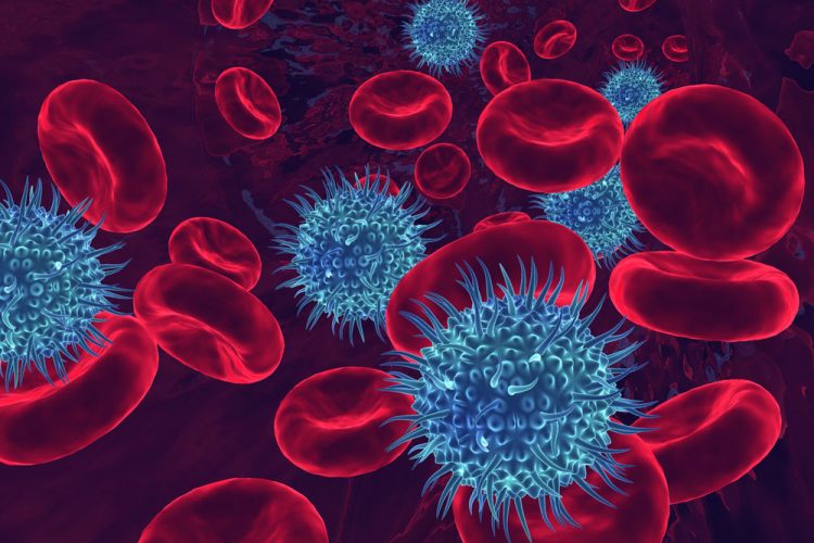
Researchers have for the first time been able to observe — live and in real-time — how the human body responds to often lethal fungal blood infections in the lung.
In the study, the fungal infection Candida albicans was introduced to mice or human models of the lung vasculature and as blood was pumped over that system, researchers recorded what happened using highly sophisticated microscopes. And what they saw surprised them.
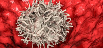
Immune cells rushed to the scene of the infection, which was expected. But then those cells swarmed, clustered and jammed up the blood vessels causing a potentially dangerous blockage, and that was a revelation, one that could lead to new treatment options for the usually deadly incursions.
This clumping of immune cells was captured in high resolution. The research study which was led by Dr Bryan Yipp, Assistant Professor in the Department of Critical Care Medicine at the Cumming School of Medicine.
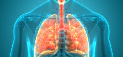
Dr Yipp and his team were interested in understanding how the lung, in particular, is involved in protecting the body from infections. “For some reason, the lung itself has a lot of the main type of immune cell, the neutrophil. We knew this, but the reasons why they are in the lung has remained mysterious,” Dr Yipp says.
As well, he says, most research has been on bacterial sepsis but they wanted to focus on fungal sepsis because it is less well understood and fungal infections are growing in number and severity. “Our initial thought was that neutrophils would capture fungi the same as they capture bacteria,” Dr Yipp says. “But they became so activated they started to clump and cluster together and actually block the bloodstream.”
The research team used sophisticated imaging techniques and novel “disease-on-chips” technology to observe the body’s response to fungal blood infections. The chips are about the size of a penny and are intended to mimic an organ system – in this case, lung vasculature.
Cumming School of Medicine researcher Dr Mark Gillrie, developed the study’s high-resolution organs-on-chips.
“We generate a pattern of an organ that we want, creating a 3D hollowed-out structure, stick it on a piece of glass, then we can introduce a lot of different cell types,” Dr Gillrie says. Astonishingly, Dr Gillrie says those cells types then have the intrinsic ability to self-organise to a certain degree, into an alveolus in the lung for example, or the blood-brain barrier in the brain.
“Our real focus is to visualise how everything actually works,” Dr Yipp says, emphasizing the imaging is everything. “These are results we would never have been able to guess at without seeing it.”
With a clearer understanding of the disease — and pictures to prove how the human body reacts to a fungal blood infection in the lung — the researchers say they are looking next to new therapeutic targets.
Dr Yipp says the goal is to find a way to block the clumping of cells without inhibiting the immune cells from capturing bugs. And the team has already had some success with anti-inflammatory drugs already on the market for asthma. “We were able to repurpose a drug that has already been approved in humans that could reduce negative consequences of infection and improve clinical outcomes,” Dr Gillrie says.
The study was recently published in CELL Host & Microbe.
Related topics
Imaging, Immunology, Microscopy, Therapeutics
Related conditions
Blood infection, Sepsis
Related organisations
University of Calgary's Cumming School of Medicine (CSM)
Related people
Dr Bryan Yipp