Breast tissue simulations address enhanced MRI delays
Posted: 25 September 2018 | Iqra Farooq (Drug Target Review) | No comments yet
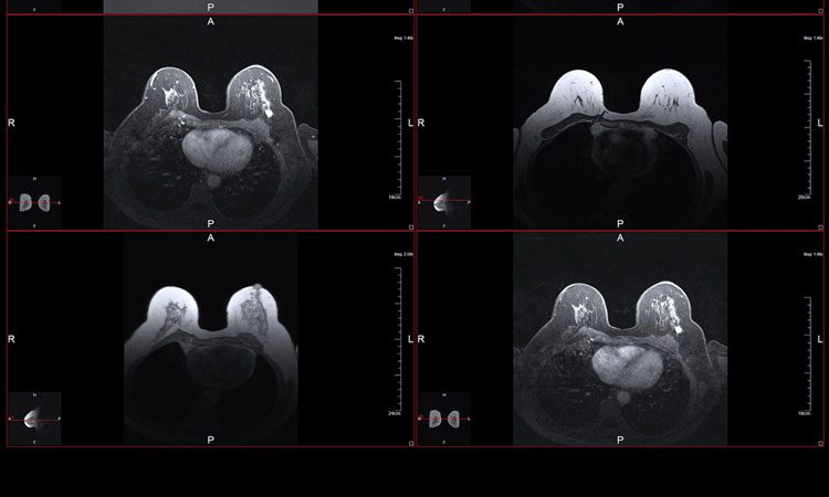
Researchers address the delay of enhanced MRI cancer detection using simulations of women’s breast tissue and higher MRI strengths…
Researchers from Purdue University argue that as no two women have the same breast tissue, MRI’s detecting and monitoring breast cancer should not treat all women the same way.
As methods of ensuring the safety of new MRIs, clinical MRIs have not been able to progress to the standard of today’s technology.
The researchers simulated how over 20 different breast tissue ratios respond to heat given off by MRIs at higher field strengths than currently available.
These simulations should show that the new technologies meet safety requirements necessary before beginning clinical trials.
The researchers even go on to predict that using knowledge about how much radiofrequency energy each breast tissue is able to handle, new techniques could use the heat generated from and target it towards tumours to kill them.
Despite clinical limitations, an annual MRI screen is recommended for women with higher than average risk of breast cancer, because it is still more sensitive than a standard mammogram.
“We’re starting to develop techniques at high field strengths that could immediately monitor how tumours respond to treatment. So we don’t want tissue heating concerns to stand in the way of improving such a powerful tool,” said Assistant Professor Joseph Rispoli.
The specific absorption rate (SAR), which indicates the amount of radiofrequency energy that will be absorbed by breast tissue from an MRI, is dependent upon how dense a woman’s breast tissue is. The more fibroglandular tissue in the breast (in comparison with breast tissue), the higher the breast density and therefore the higher the SAR.
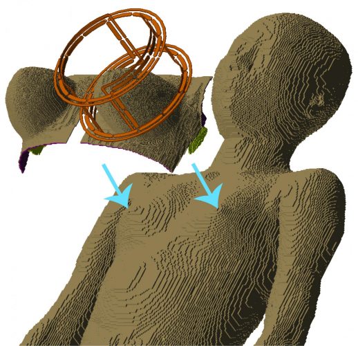
There are far fewer computer models of the female body compared to the male body, making it difficult for researchers to predict that a new MRI technique would be safe for all women’s breast tissue. Credit: Purdue University image/Xin Li.
“In hospitals, MRI field strengths are currently up to 3 tesla, tesla being the unit we use for measuring magnetic field strength,” Prof Rispoli said. “Many techniques would be far superior at 7 tesla. This would come at a fivefold increase in SAR, but also double the MRI sensitivity.”
The researchers used full body ‘phantoms’ (computer models) of the female body to test their techniques. They fused 36 breast phantoms at various densities with two full body female phantoms. They then simulated the behaviour of each fused phantom in response to their MRI at the higher rate of MRI field strength.
“We want to facilitate the most cutting-edge of breast MRI techniques at any site in the world,” Prof Rispoli said. “Ultimately, a woman will be able to go in, have a fast low-power anatomical MRI scan, and then the computer could quickly simulate on-the-fly what the SAR would be in that patient.”
The findings of the study were published in the journal Magnetic Resonance in Medicine.
Related topics
Disease Research, Imaging, Oncology, Research & Development, Screening
Related conditions
Breast cancer
Related organisations
Purdue University
Related people
Assistant Professor Joseph Rispoli