Local gyrification index, which measures cortical folding in the brain, could be a novel neuroimaging marker for major depressive disorder.

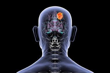
In appearance, the human brain’s outermost layer, called the cortex, is a maze of tissue folds. The peaks or raised surfaces of these folds, called gyri, play an important role in the proper functioning of the brain. Improper gyrification—or the development of gyri—has been implicated in various neurological disorders, one of them being the debilitating and widespread mental illness, major depressive disorder (MDD).
In a positive turn of events, researchers led by Professor Byung-Joo Ham and Associate Professor Kyu-Man Han from Korea University College of Medicine, South Korea, have reported the successful identification of a neuroimaging-based biomarker for MDD in a recent study published in Psychological Medicine.
Although prior studies have shown that abnormal cortical folding patterns are associated with MDD, a reliable indicator has so far remained out of reach.
Talking about the unique finding that sets apart their study from the previous ones, Ham explained, “Our first-of-its-kind study investigated the association of MDD with the local gyrification index or LGI of multiple cortical regions at the whole-brain level and the association of LGI with the clinical characteristics of MDD.”
But what indeed is local gyrification index (LGI)?
LGI is a measurement of cortical folding that is derived from brain scans as a ratio of the curved and smoothed surfaces of the cortex in a region of interest. The researchers compared the LGI values from multiple cortical regions in the brain of patients with MDD with those of healthy individuals. The neuroimaging data used to compare and analyse both groups were obtained from magnetic resonance imaging scans.

Local gyrification index may be a relatively stable neuroimaging marker for establishing the predisposition to major depressive disorder. (Credit: Kyu-Man Han and Byung-Joo Ham)[/caption]
LGI values from multiple cortical regions in the brain of patients with MDD showed hypo-gyrification- a condition characterised by decreased cortical folding, when compared with healthy individuals. They found that patients with MDD showed significantly lower LGI values in 7 out of the 66 cortical regions assessed (in both hemispheres of the brain), which included the prefrontal cortex, anterior cingulate cortex, insula, and several temporal and parietal regions. Notably, the most significant hypo-gyrification was observed in the left pars triangularis of patients with MDD. These findings are nothing short of a breakthrough in MDD research!
When asked to share his thoughts about the study’s results, Han asserts that there is more to their findings than what meets the eye. “The cortical regions that we assessed in our study have been previously shown to affect emotional regulation. This means that abnormal cortical folding patterns may be associated with the dysfunction of neural circuits involved in emotional regulation, thus contributing to the pathophysiology of MDD.”
The study’s findings firmly establish LGI as a relatively stable neuroimaging marker for MDD, when compared with previously identified biomarkers. This is because LGI values reflect the long-drawn developmental process of gyrification that is not spontaneously affected by an individuals’ state during the measurement process. It is also worth highlighting the robustness of this study, given that it involved a larger sample size of participants, which gives it an edge over similar studies conducted previously.
Interestingly, the researchers noted that the clinical characteristics of MDD, including the recurrence and duration of illness in patients, were associated with increased gyrification in several occipital and temporal cortical regions. However, they did not observe any significant difference in the LGI values in these regions between the patient and control groups.
With the head start given by this study, future research could explore the genetic factors that predispose individuals to abnormal cortical folding patterns, and, in turn, MDD. The study can also serve as a roadmap for the selection of cortical regions as targets for medical treatments aimed at reducing the symptoms of this condition.