3D tumour models enable scientists to offer better treatments and improve survival outcomes for complex conditions like breast cancer.
Scientists from University of Waterloo have created a method which produces better three-dimensional (3D) models of complex cancers. The team combined innovative bioprinting techniques with synthetic structures or microfluidic chips which will enable lab researchers to more accurately understand heterogeneous tumours - tumours with more than one kind of cancer cell, often dispersed in unpredictable patterns.
Traditionally, medical practitioners would biopsy a patient’s tumour, extract cells, and then grow them in flat petri dishes in a lab. Nafiseh Moghimi, Applied Mathematics post-doctoral researcher and lead author of the study said: “For fifty years, this was how biologists understood tumours.” She continued: “But a decade ago, repeated treatment failures in human trials made scientists realize that a 2D model does not capture the real tumour structure inside the body.”
The team addressed this issue by creating a 3D model that simulates the complexity of the tumour and its surrounding environment.
“We are creating something that is very, very new in Canada. Maybe just a couple of labs are doing something even close to this research,” Moghimi said.
The research took place under the supervision of Applied Mathematics Professor Mohammad Kohandel in the Mathematical Medicine Lab. To begin, the team engineered polymer “microfluidic chips”, miniscule structures etched with channels that mimic blood flow and other fluids surrounding a patient’s tumour.
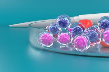
Then, the team grew multiple types of cancer cells and suspended these cell cultures in their own customised bioink. The bioink contained gelatine, alginate, and other nutrients intended to keep the cells cultures alive.
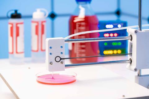
The third step was to use an extrusion bioprinter, a device that resembles a 3D printer but for organic material, to layer the different types of cancer cells onto the prepared microfluidic chips.
This resulted in a living, 3D model of complex cancers that researchers can then use to test different modes of treatment, like multiple chemotherapy drugs.
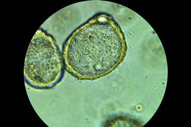
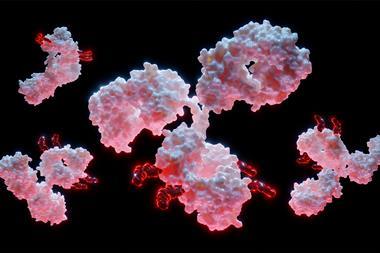
Moghimi and her team are especially interested in creating complex models of breast cancer. After skin cancer, breast cancer is the most common cancer diagnosed in women and is difficult to treat because it presents as complex tumours containing multiple types of cells when it metastasises. Biopsies do not accurately represent the entire tumour and can result in ineffective treatment plans and undesirable outcomes. These 3D-printed tumour models offer promise for quicker, cheaper, and less painful treatments for conditions like late-stage breast cancer.
This research is published in Nature.