A new method for a fragmentation-based identification of lipids could enable the study of cancer cells in detail not seen before.
Led by the University of Surrey, scientists have developed and validated an untargeted single-cell lipidomics method for a fragmentation-based identification of lipids. In the study, single live cancer cells were sampled and the fatty lipid compounds inside them were measured to investigate how they transformed in response to changes in their environment. This work was conducted with partners at GSK and UCL, as well as Yokogawa, who developed new equipment for the research.
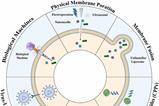
Understanding the variation of lipids in single cells will aid further research in many biological processes, as lipids have significant roles in biophysical, energy storage, structural and signalling functions.
Dr Johanna Von Gerichten, from the School of Chemistry and Chemical Engineering at the University of Surrey, explained: “The trouble with cancer cells is that no two are alike. That makes it harder to design good treatment, because some cells will always resist treatment more than others…Yet it has always proven tricky to study live cells after they have been removed from their natural environment, in enough detail to truly understand their make-up.”
She added: “That is why it is so exciting to be able to sample live cells under a microscope and study their fatty contents one by one.”
The most common pancreatic cancer with the poorest prognosis is pancreatic ductal adenocarcinoma (PDAC). However, analysing single cells presents multiple difficulties because of the low abundance of analytes in one cell. To overcome this challenge, individual pancreatic cancer cells were lifted from a glass culture dish using Yokogawa’s Single Cellome System SS2000 ™, which extracts single live cells using tubes measuring 10 µm across.
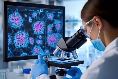
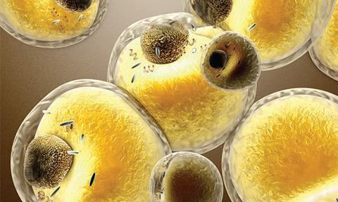
The researchers stained the cells with fluorescent dye to monitor lipid droplets throughout the experiment. Working with partners at Sciex, the team then developed a new method using a mass spectrometer to fragment the lipids in the cells to inform them of their composition. It was shown that different cells had very different lipid profiles, and that lipids in the cells changed in response to what was going on around them.
Dr Carla Newman, Associate Director, Cellular Imaging and Dynamics at GSK commented: “Our new method paves the way for studying cancer cells in detail we’ve never seen before…One day, we might be able to see how individual cancer cells communicate with their neighbours. That could unlock new, more targeted treatments.”
The study was published in Analytical Chemistry.