Mini-colons reveal complex process of tumorigenesis ex vivo
Posted: 25 April 2024 | Drug Target Review | No comments yet
The mini-colons are topobiologically complex, can be induced to develop tumours in targeted areas and reduce the use of animal models.
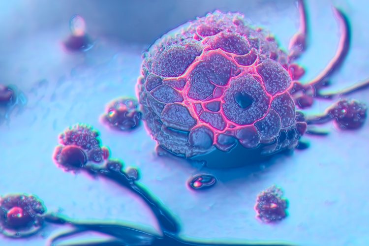
Scientists part of the Matthias Lütolf research group at EPFL have developed miniature colon tissues that can simulate the complex process of tumorigenesis outside of the body with high fidelity. Created by combining microfabrication and tissue engineering techniques, these colon tissues give rise to tumours that closely resemble those found in vivo. This breakthrough will enable a better understanding of the processes underlying cancer initiation, progression and response to treatment.
The requirement for more sophisticated and realistic models to study tumour development is critical. Research has relied on animal models, simplified cell culture methods, or organoids which are valuable but cannot fully capture the interplay of factors involved in tumour development.
The mini-colons are topobiologically complex, replicating the physical structure of colon tissue, including its distinctive crypt-and-lumen architecture, and mimicking the cellular diversity present in the actual colon tissue during healthy and diseased states. Another important feature of the mini-colons is that they can be induced to develop tumours “at will” and in targeted areas, which mimics the localised onset of colorectal cancer. The researchers were able to turn inducible oncogenic genes on using optogenetics.
Integrating a blue-light-responsive system into the mini-colons meant they underwent controlled oncogenic mutations, which can reveal tumour evolution with a high level of detail. Dr Matthias Lütolf, the founding director of Roche’s new Institute of Human Biology, explained: “In essence, we used light to trigger tumorigenesis by turning on oncogenic driver mutations in a spatiotemporally controlled manner in healthy bioengineered colon epithelial organoids.”
Triggering these genetic changes with light in the miniature colons also provides a powerful tool to study the dynamic processes of tumour development and the cellular response to these mutations in real-time, enabling new possibilities for dissecting the molecular and cellular mechanisms of cancer. The researchers manipulated genetic and environmental conditions to replicate and observe a range of tumour behaviours in the mini-colons, and even identified key factors influencing cancer progression. For instance, the found the GPX2 protein which associated with stem cell characteristics and tumour growth.
This study means that scientists can explore the underlying mechanisms of colorectal cancer and test potential therapies, particularly when applied to human patient-derived tissues. The mini-colons also reduce our use of animal models.
This study was published in Nature.
Related topics
Cancer research, Drug Targets, Ex Vivo, Optogenetics, Organoids
Related conditions
Cancer Research, Colorectal cancer
Related organisations
EPFL
Related people
Dr Matthias Lütolf (EPFL)