

UV light microscope proves useful diagnostic tool
Posted: 6 December 2017 | Drug Target Review | 1 comment
Recent study shows how a microscope using ultraviolet (UV) light to illuminate samples enables pathologists to assess high-resolution images of biopsies and other fresh tissue samples for disease, quickly and effectively.
This approach provides clear images within minutes, eradicating the need for time-consuming preparation of conventional slides and without destroying the tissue. A recent study involving UC Davis and the University of California has demonstrated how this UV technology could improve the speed and efficiency of patient care and medical research nationwide.
Microscopy with UV surface excitation
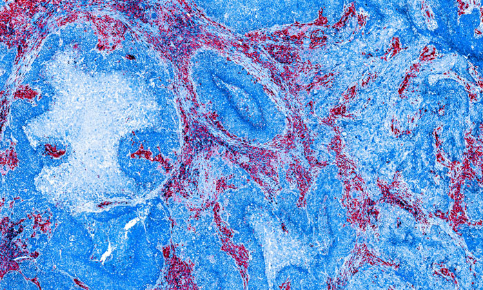
The technology – known as microscopy with UV surface excitation, or MUSE – uses ultraviolet light at wavelengths below the 300 nanometre range to penetrate the surface of tissue samples by only a few microns (about the same thickness of tissue slices on traditional microscope slides.) The phenomenon was originally described by Stavros Demos, one of the co-authors, who is now at the University of Rochester.
Samples that have been stained with eosin or other standard dyes to highlight important features – such as nuclei, cytoplasm and extracellular components – produce signals from the UV excitation that are bright enough to be detected by conventional colour cameras using sub-second exposure times. The process allows for rapid imaging of large areas and immediate interpretation.
Biopsy application
Commenting on the benefits of the new microscope technology, Richard Levenson, Professor and Vice Chair for Strategic Technologies in the Department of Pathology and Laboratory Medicine at UC Davis and senior author of the study, said: “MUSE eliminates any need for conventional tissue processing with formalin fixation, paraffin embedding or thin-sectioning. It doesn’t require lasers, confocal, multiphoton or optical coherence tomography instrumentation, and the simple technology makes it well suited for deployment wherever biopsies are obtained and evaluated,” he said.
MUSE’s ability to quickly gather high-resolution images without consuming the tissue is a particularly important feature.
“It has become increasingly important to submit relevant portion of often tiny tissue samples for DNA and other molecular functional tests,” he said. “Making sure that the submitted material actually contains tumour in sufficient quantity is not always easy and sometimes just preparing conventional microscope slices can consume most of or even all of small specimens.
MUSE is important because it quickly provides images from fresh tissue without exhausting the sample.”
The ability to obtain instant, high-resolution, full-colour images for histology, pathology or toxicology studies is also useful for basic scientists who want to assess tissue samples from experimental animal models at the laboratory bench.
Related topics
Imaging, Oncology, Technology
Related organisations
UC Davis
Related people
Richard Levenson, Stavros Demos

Hello, At first i wanted to thanks for the article, it helped me a lot. And secondly i wanted to know about the tissue that it´s shown. What tissue it is?