RhoA protein function identified in nerve cell development
Posted: 4 November 2019 | Drug Target Review | No comments yet
A recent study has revealed new findings about nerve cell development that could help to facilitate future treatment options for spinal cord injuries.
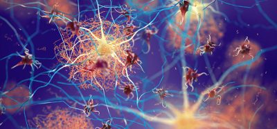
During embryonic development, nerve cells form long, thin extensions called axons, which they use to wire up the complex network of the brain. Scientists from the German Center for Neurodegenerative Diseases (DZNE) in Bonn have now identified a protein that regulates the growth of these extensions, called RhoA, by pulling a brake. Long term, these findings could help to develop new approaches for the treatment of spinal cord injuries.
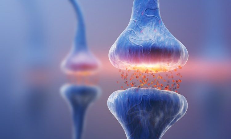
Neurons transmit electrical signals in a clearly defined direction and are said to be ‘polarised’. Each neuron receives signals and forwards them via a long extension, the so-called axon, to the next cell. In humans, axons in the spinal cord can grow to become one metre in length. Scientists in Bonn questioned whether it is possible to restimulate this impressive growth potential after a spinal cord injury.
“To answer this question, we first need to better understand the molecular processes underpinning embryonic development,” said Professor Frank Bradke, group leader at DZNE’s Bonn site and head of the study. He and his colleagues have now come a step closer to this goal by investigating neuronal growth in mice and cell culture.
The workhorse RhoA protein
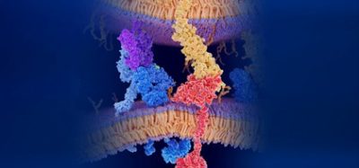
Their work has focused on a protein called RhoA – a jack-of-all-trades among molecules, which interacts with many protein partners and has different functions in a wide variety of cells. However, until now its precise function in neurons had not been determined.
“For a long time it was thought that RhoA would determine the polarity of the neuron and thus select the position of axon formation in the cell,” explained Bradke. The current study, however, shows that this is not the case: RhoA has little to do with cell polarity and axon specification. Rather, RhoA comes into play only after the axon is formed and regulates its extension via a molecular cascade.
This insight could be important for new therapies. “Manipulating the RhoA signalling pathway should thus only affect the growth of the nerve fibres without disturbing the cell’s internal organisation,” said Bradke.
Bradke and his colleagues demonstrated that RhoA activates a molecular signalling pathway that directly targets neurons’ cytoskeletons. RhoA restrains axonal elongation by restricting the advancement of so-called microtubules – cytoskeletal building blocks necessary for axon stabilisation – into the growth zone of the axon.
“In embryonic development, such a growth brake is probably necessary to coordinate different developmental processes. An accurate understanding of its molecular basis could now help to advance research on spinal cord regeneration after injury. To this end, the brake would need to be released,” says Dr Sebastian Dupraz, the lead author of the study and a postdoctoral fellow in Bradke’s lab. “The molecular cascade we have identified directly influences the cytoskeleton of the axon and thus provides a good starting point for therapeutic strategies.”
The study is published in the journal Current Biology.
Related topics
Disease Research, Drug Targets, Neurons, Protein, Regenerative Medicine, Targets
Related conditions
Spinal cord injuries
Related organisations
German Center for Neurodegenerative Diseases
Related people
Dr Sebastian Dupraz, Professor Frank Bradke