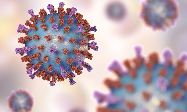
Structure of key pneumonia virus enzyme uncovered
Posted: 10 December 2019 | Rachael Harper (Drug Target Review) | No comments yet
Scientists have used cryo-electron microscopy to clarify the structure of one of the key components of RSV and HMPV, which could lead to new therapies for the viruses.


Scientists have found a potential new route to disabling respiratory syncytial virus (RSV) and human metapneumovirus (HMPV) after elucidating the structure of one of its key components.
The scientists were from Nanyang Technological University, Singapore (NTU Singapore), led by Dr Julien Lescar, an Associate Professor at NTU’s School of Biological Sciences
Analysis of these model structures revealed key sites for molecules to interact at, offering new targets”
RSV and HMPV are two closely related viruses causing severe and life-threatening respiratory diseases such as pneumonia and bronchiolitis in anyone with a weak immune system. HMPV and RSV commandeer human cells’ machinery to make copies of themselves. To initiate the process, special proteins released by the virus interact with each other to make distinct protein complexes.
Dr Lescar his team have used cryo-electron microscopy to image the molecular structure of one of these large complexes, an enzyme called HMPV L:P polymerase.
The images captured the enzyme at a resolution of 3.7 Angstrom, or 0.37 nanometres. Based on these two-dimensional pictures, the team then built three-dimensional computer models of the proteins’ L:P molecular structures.
Analysis of these model structures revealed key sites for molecules to interact at, offering new targets for designing antiviral molecules against both viruses.
Dr Lescar said with this detailed structural knowledge, researchers can now hope to develop inhibitors that disrupt the enzymatic activities of HPMV L:P protein and potentially block infection by the virus.
“We hope that our work will help researchers in pharma and academia around the world to design much-needed therapies for difficult viral infections that often lead to antibiotic-resistant bacterial infections,” said Dr Lescar.
Since the HMPV proteins they studied are essentially unchanged through evolution and very similar to those of RSV and other virus species belonging to the Pneumorivridae family, the scientists have said they hope that inhibitors developed against HPMV could also work against a broad spectrum of viruses involved in respiratory diseases and inform similar quests against other viral diseases.
The study was published in Nature.
Related topics
Disease Research, Drug Targets, Enzymes, Imaging, Microscopy, Therapeutics
Related conditions
Bronchiectasis, pneumonia
Related organisations
Nanyang Technological University (NTU)
Related people
Dr Julien Lescar


