Scientists reveal structure of norovirus using cryo-EM
Posted: 1 April 2020 | Victoria Rees (Drug Target Review) | No comments yet
Researchers captured 13,000 images of a mouse norovirus using an electron microscope and compiled the images to reveal the structure of the virus.
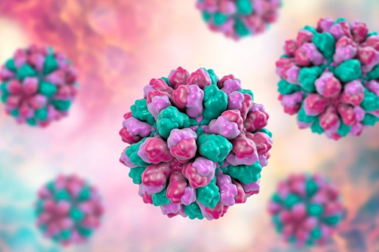
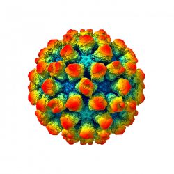
This is an image of the murine norovirus captured by cryo-electron microscopy [credit: University of Leeds].
A group of researchers has combined images taken by an electron microscope (EM) to compile a detailed image of the structure of the norovirus.
The study was conducted at the University of Leeds, UK, and the researchers took 13,000 separate images using high-resolution cryo-EM to produce their findings. For safety reasons, the researchers used a mouse norovirus in their study, which closely resembles human noroviruses.
The scientists explain that their image shows bump-like protrusions on the outside of the virus capsid, the protein shell that holds the genome of the virus.
Joseph Snowden, a PhD researcher from the Astbury Centre for Structural Molecular Biology at the University of Leeds, said it shows that the protein casing does not keep a fixed shape – as the protrusions on the surface would extend, retract and rotate.
According to the scientists, this dynamic shape-shifting may hold clues as to why the noroviruses are such potent pathogens.
To identify subtle changes to the structure of these protein shells, the scientists processed the data from the images with a super computer, using a method called ‘focused classification’.
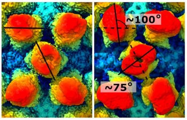
The image shows how the protrusions on the exterior of the virus change shape [credit: University of Leeds].
Snowden said: “The constant changing or morphing of the virus shell may enable it to confuse the body’s defence systems. Immune systems work on the basis of molecular shape. The body’s defences will produce proteins that bind to pathogens to prevent them from infecting host cells. However, if the shape of the virus is constantly changing, the body’s defence systems may be unable to act efficiently.”
The protrusions seemed able to move independently or in a co-ordinated fashion and the scientists believe this may enable the virus to prime itself to become ready for infection depending on where it is, ie, if it is in the digestive tract of a host organism.
Dr Morgan Herod, one of the senior authors on this study, also from the University of Leeds, hopes that a greater understanding of the structure of the norovirus may help with vaccine development, which so far has been unsuccessful.
“The University of Leeds has a strong track record in the use of virus like particles (VLPs) to create vaccine candidates, for example against polio. VLPs are harmless proteins modelled on the structure of a virus’s casing and they fool the immune system into thinking the body is under attack, prompting an immune response… Our research shows that the capsid or protein shell of norovirus is dynamic – and perhaps we have to look at VLPs that are better able to mimic this aspect of norovirus structure,” said Dr Herod.
The study was published in PLOS Biology.
Related topics
Imaging, Microscopy, Research & Development, Structural Biology
Related conditions
norovirus
Related organisations
Leeds University
Related people
Dr Morgan Herod, Joseph Snowden