Novel imaging technique captures multiple tissues in 3D
Posted: 30 November 2020 | Victoria Rees (Drug Target Review) | No comments yet
A new imaging method called FLASH can provide a visualisation of several tissue types in a 3D format, its developers say.
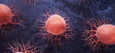
A team has developed a new imaging technique that allows simultaneous visualisation of multiple tissue types in three-dimensions (3D). The researchers say the method provides one of the most comprehensive views of conditions like cancer to date. The approach was developed at the Francis Crick Institute, UK.
The research team set out to address problems with existing histology techniques that prevent a holistic view of diseases like cancer. According to the scientists, most imaging methods require ultra-thin samples of just a few micrometres thick, which may not be reflective of the rest of the sample and cannot provide insight into the spatial interactions between different cell types.
Over a period of about two years, the team trialled different combinations of reagents, techniques and timings, until they managed to find a process that allowed them to capture the detailed 3D images, while leaving the sample or organ intact for further analysis. They named their new technique fast light microscopic analysis of antibody stained whole organs (FLASH).
Biomarkers are redefining how precision therapies are discovered, validated and delivered.
This exclusive expert-led report reveals how leading teams are using biomarker science to drive faster insights, cleaner data and more targeted treatments – from discovery to diagnostics.
Inside the report:
- How leading organisations are reshaping strategy with biomarker-led approaches
- Better tools for real-time decision-making – turning complex data into faster insights
- Global standardisation and assay sensitivity – what it takes to scale across networks
Discover how biomarker science is addressing the biggest hurdles in drug discovery, translational research and precision medicine – access your free copy today
Hendrik Messal, first author and postdoctoral fellow at the Netherlands Cancer Institute, said: “The concept of 3D imagery is fast evolving and researchers are always testing the boundaries of what we can see using the latest technologies and techniques. FLASH is a fast and easy method that can be quickly adopted by researchers familiar with light microscopy techniques and importantly, it does not damage the tissue which allows for additional diagnostic tests.”
Video: 3D FLASH image of a mouse lung, showing bronchiole and arteriole (magenta), clara cells (green) and alveolar typeII cells (cyan)
The team showed how the technique can be used to examine the architecture of pancreatic tumours and predict how aggressive the cancer would become.
“Thin histological sections might miss some clues of cancer biology,” said Jorge Almagro, first author and PhD student at the Crick. “Examining a whole sample provides researchers with a panoramic view of disease – which cell types are present and how they are interacting.”
Video: 3D FLASH of mammary epithelium showing luminal cells (pink), myoepithelial cells (yellow) and basal lamina defining adipocytes
The research team say they are continuing to refine this method and adjust it to suit different tissue types and research questions.
Details of the new imaging method are published in Nature Protocols.
Related topics
Disease Research, Imaging, Microscopy, Oncology
Related conditions
Cancer, Pancreatic cancer
Related organisations
Francis Crick Institute, Netherlands Cancer Institute
Related people
Hendrik Messal, Jorge Almagro