Novel spectroscopic microscope images biological molecular conformations
Posted: 9 March 2021 | Victoria Rees (Drug Target Review) | No comments yet
A spectroscopic microscope has been developed by researchers to gather data on biological conformations faster and more accurately.
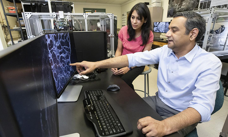
Rohit Bhargava and graduate student Yamuna Phal analysing a microscopic image of solid tissue. This photo was taken in February 2020 [credit: L Brian Stauffer, University of Illinois Urbana-Champaign].
Researchers have developed a spectroscopic microscope to enable optical measurements of molecular conformations and orientations in biological samples. The novel measurement technique allows researchers to image biological samples at the microscopic level faster and more accurately. The method was developed at the University of Illinois Urbana-Champaign, US.
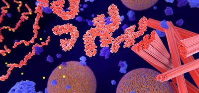
“This project is about bringing the study of molecular chirality into the microscopic domain,” said Professor Rohit Bhargava, one of the lead researchers. According to the researchers, molecular chirality refers to the spatial orientation of atoms in molecules or multimolecule assemblies. In biological systems, one molecule may elicit a cellular response, while its mirror image could be inactive or even toxic. While vibrational circular dichroism (VCD) can be employed to help determine a molecule’s chemical structure and orientation, these measurements are time-intensive and could not previously be used to image complex biological systems or solid tissues samples.
The novel infrared microscope makes imaging of biomolecule chirality possible by accelerating both the acquisition time and improving the signal-to-noise ratio of traditional VCD techniques, the researchers say.
“When you send light down a microscope from a spectrometer, you are essentially throwing away a lot of it,” Bhargava said. “For VCD measurements, you also have to send it through a photoelastic modulator, which changes the polarisation of light to left- or right-handed. At that point, you do not have a lot of light left, which means you have to average your signal for a long time to see just one pixel within an image.”
The researchers achieved rapid and concurrent infrared and VCD measurements by building on the framework of their high performance discrete frequency infrared imaging microscope. Instead of employing a traditional thermal light source, the instrument is built around a quantum cascade laser.
“The laser source motivated the whole design,” said Yamuna Phal, a graduate student researcher. “The QCL source has higher power, which means we can acquire faster measurements. Previously, you could only perform VCD on liquid samples, but we can image solid tissues as well. This was never attempted before because it takes so long to acquire VCD signals in the first place.”
The study was published in Analytical Chemistry.
Related topics
Imaging, Microscopy, Molecular Targets
Related organisations
University of Illinois Urbana-Champaign
Related people
Professor Rohit Bhargava, Yamuna Phal