Atomic force microscopy used to study endonuclease digestion of DNA nanostructures
Posted: 2 March 2021 | Victoria Rees (Drug Target Review) | No comments yet
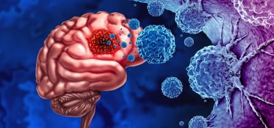
A team of researchers have used microscopy techniques to monitor DNA degradation and anticancer drug release from nanostructures.

A team of researchers from Aalto University, the University of Jyväskylä, both Finland, Ludwig-Maximilian-Universität München and Universität Paderborn, both Germany, have discovered a way to study the endonuclease-driven digestion of drug-loaded DNA nanostructures in real time.
According to the researchers, DNA nanotechnology has developed rapidly during recent years and enabled the construction of increasingly complex nanostructures. DNA nanostructures, such as DNA origami, serve as an excellent foundation for nanocarrier-based drug delivery applications. Although the stability of such nanostructures under physiological conditions can be improved, little is known about their digestion by endonucleases, which are responsible for destroying foreign DNA in our bodies.
The researchers’ previous experiments used high-speed atomic force microscopy to show that the design of DNA origami plays a role in how quickly they break apart in an endonuclease-rich environment. While they could follow the digestion process at a single-structure level, the approach was limited to two-dimensional (2D) origami shapes deposited on a microscope substrate.
Now the group has monitored DNA degradation and the subsequent anticancer drug doxorubicin (Dox) release from the structures. The drug bonds between DNA base pairs.
“We observed both the digestion and drug release profiles as the drug is released upon DNA fragmentation by nucleases and importantly, in the solution phase. With this method we can actually see the collective behaviour of all the nanostructures when they are floating freely in liquid,” said Adjunct Professor Veikko Linko from Aalto University, who led the study.
According to the researchers, the structural integrity was verified by atomic force microscopy and transmission electron microscopy.
“It seems the digestion happens differently on substrates and in solution and by combining these two types of information, we can better understand how the nanostructures are digested by nucleases in the bloodstream. Moreover, we showed that the drug release profiles were closely linked to the digestion profiles and a wide range of drug doses could be achieved simply by changing the shape or geometry of the DNA nanostructure,” explained doctoral student Heini Ijäs, the main author of the research.
As the team investigated the binding of Dox to the DNA structures in great detail, they discovered that the majority of previous studies have vastly overestimated the Dox loading capacity of DNA origami.
“The anticancer effects of Dox-equipped DNA nanostructures have been reported in many publications, but it seems these effects may have been mainly caused by free or aggregated Dox molecules, not by the drug-loaded DNA motifs. We believe this type of information is crucial for the development of safe and more effective drug delivery systems and brings us one step closer to real-world DNA-based biomedical applications,” said Ijäs.
The research was published in Nucleic Acids Research.
Related topics
DNA, Imaging, Label-Free, Microscopy, Nanomedicine
Related organisations
Aalto University, Ludwig-Maximilian-Universität München, Universität Paderborn, University of Jyväskylä
Related people
Adjunct Professor Veikko Linko, Heini Ijäs