Clearer imaging approach captures images of blood vessels
Posted: 2 March 2022 | Ria Kakkad (Drug Target Review) | No comments yet
Researchers have developed a new imaging approach that can capture blood vessel images at different spatial scales.
Researchers from Johns Hopkins Medicine, US have developed a new approach that can accelerate imaging-based research by allowing investigators to capture images of blood vessels at different spatial scales. The study, which was recently published in Nature Methods, tested the method called “VascuViz,” in mouse tissues and included a quick-setting polymer mixture to fill blood vessels and make them visible in multiple imaging techniques.
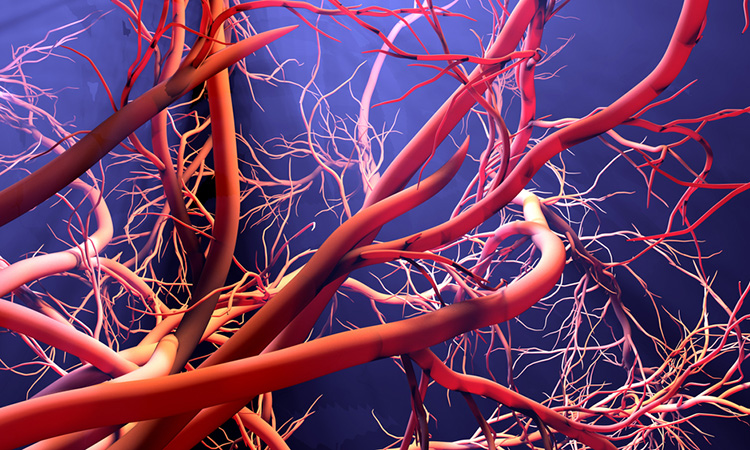
A still from the visual demonstration of the result of the VascuViz imaging pipeline.
[Credit: Johns Hopkins Medicine].
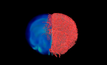
The approach enabled researchers to visualise the structure of a tissue’s vasculature, which in conjunction with detailed mathematical models or complementary images of other tissue elements can clarify the complex role of blood flow in health and diseases. The team say the combined images of the blood vessels can enhance the study of the biology of diseases that involve abnormalities in blood flow and advance information about the structures and functions of tissues throughout the body.
Usual methods to study the role of blood vessels include magnetic resonance imaging (MRI), computerised tomography (CT) and microscopy. Although these methods are useful for understanding how tissues develop disease and respond to treatment, agents used to make a blood vessel visible to one imaging method can make it invisible on other tools. This reduces the amount of data researchers can collect from a single sample.
VascuViz made the structure of the largest arteries to the smallest microvasculature visible to a variety of imaging tools, which allowed researchers to develop a multi-layered understanding of blood vessels and related tissue components with less time and effort. The development of VascuViz can be useful in creating computerised visualisations of complex biological systems, such as the circulatory system and is a feature of the growing field of “image-based” vascular systems biology.
“Now, rather than using an approximation, we can more precisely estimate features like blood flow in actual blood vessels and combine it with complementary information, such as cell density,” said Dr Akanksha Bhargava. To do this, VascuViz-based measurements are entered into computer simulations of blood flow, such as the cancer models Bhargava studied.
To create VascuViz, Bhargava tested several combinations of existing imaging agents and their suitability for different imaging methods. After multiple iterations, she found that a CT contrast agent and a fluorescently labelled MRI contrast agent could be combined to create a compound that makes the macro- and microvasculature simultaneously visible when imaging with MRI, CT and optical imaging techniques without interference.
With the compound working in test tubes, the researchers then tested it in a variety of mouse tissues, perfusing it through the vascular system of breast cancer models, leg muscles, the brain and kidney tissues. The resulting images of the tissues acquired with MRI, CT and optical microscopy were then combined to create three-dimensional visualisations of the vasculature and associated components comprising these disease model and organ systems.
Due to VascuViz’s affordability, the team hope it is globally adopted by scientists to help shed new light on different diseases involving the vasculature.
Related topics
Computerised Tomography (CT), Drug Discovery Processes, Imaging, Magnetic resonance images (MRI), Microscopy
Related organisations
Johns Hopkins Medicine
Related people
Dr Akanksha Bhargava