Retinal cell map could advance precise cell and gene therapies for retinal diseases
Posted: 10 May 2022 | Ria Kakkad (Drug Target Review) | No comments yet
Researchers have identified distinct differences among the cells comprising a tissue in the retina, findings that could help develop precise therapies for retinal diseases.

In a new study, researchers form the US National Eye Institute (NEI) have identified distinct differences among the cells comprising a tissue in the retina that is vital to human visual perception. The scientists discovered five subpopulations of retinal pigment epithelium (RPE), a layer of tissue that nourishes and supports the retina’s light-sensing photoreceptors. Using artificial intelligence (AI), the researchers analysed images of RPE at single-cell resolution to create a reference map that locates each subpopulation within the eye. The ground-breaking research, which was recently published in Proceedings of the National Academy of Science, could help find more precise cell and gene therapies for retinal diseases.
Age and disease can cause metabolic changes in RPE cells that can lead to photoreceptor degeneration. The impact on vision from these RPE changes varies dramatically by severity and where the RPE cells reside within the retina. For example, late-onset retinal degeneration (L-ORD) affects mostly peripheral retina and, therefore, peripheral vision. Age-related macular degeneration (AMD), a leading cause of vision loss, primarily affects RPE cells in the macula, which is crucial for central vision.
The researchers aimed to determine if there are different RPE subpopulations that might explain the wide spectrum of retinal disease phenotypes. They used AI to analyse RPE cell morphometry, the external shape and dimensions of each cell. They trained a computer using fluorescently labelled images of RPE to analyse the entire human RPE monolayer from nine cadaver donors with no history of significant eye disease.
Morphometry features were calculated for each RPE cell – on average, about 2.8 million cells per donor; 47.6 million cells were analysed in total. The algorithm assessed each cell’s area, aspect ratio (width to height), hexagonality, and number of neighbours. Previous studies had suggested that RPE function is tied to the tightness of cellular junctions; the more crowded, the better for indicating cellular health.
Based on morphometry, they identified five distinct RPE cell subpopulations, referred to as P1-P5, organised in concentric circles around the fovea, which is the centre of the macula and the most light-sensitive region of the retina. Compared to RPE in the periphery, foveal RPE tend to be perfectly hexagonal and more compactly situated, with higher numbers of neighbouring cells.
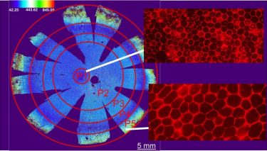
Using artificial intelligence, the researchers analysed RPE from nine donor and identified five subpopulations of RPE. These populations are along a spectrum in terms of cell area, aspect ratio, hexagonality and number of neighbours. Foveal RPE (P1) required for central vision tend to be perfect hexagons and are tightly packed together. Peripheral RPE (P5) are less perfect hexagons and are more spread out
[Credit: Dr Davide Ortolan].
Next, they analysed RPE from cadavers with AMD. Foveal (P1) RPE tended to be absent due to disease damage, and the differences among cells in the P2-P5 subpopulations were not statistically significant. Overall, the AMD RPE subpopulations tended to be elongated relative to RPE cells not affected by AMD.
To further test the hypothesis that different retinal degenerations affect specific RPE subpopulations, they analysed ultrawide-field fundus autofluorescence images from patients affected by chloridaemia, L-ORD, or a retinal degeneration with no identified molecular cause. While these studies were conducted at a single point in time, they still demonstrated that different RPE subpopulations are vulnerable to different types of retinal degenerative diseases.
Age-related morphometric changes also may appear in some RPE subpopulations before they’re detectable in others. These finding could help inform future studies using non-invasive imaging technologies, such as adaptive optics, which resolve retinal cells in unprecedented detail and could potentially be used to predict changes in RPE health in living patients.
Related topics
Artificial Intelligence, Disease Research, Gene Therapy, Imaging, Stem Cells
Related conditions
age-related macular degeneration (AMD), blindness, Retinal disease
Related organisations
US National Eye Institute (NEI)