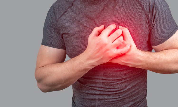
New discovery could open ways to repair damage in the human body after stroke or heart attack
Posted: 6 May 2022 | Ria Kakkad (Drug Target Review) | No comments yet
New data from high-resolution imaging has revealed the origin of circulatory system during development, a discovery that could help advance therapeutics for various vasculature-related pathologies.
Using high-resolution, time-lapse imaging and a technique called transcriptional profiling to understand relationships between cells, scientists at the Fralin Biomedical Research Institute at Virginia Tech Carilion School of Medicine and Research Institute (VTC), US found separate but closely connected cell types working together to build blood and other circulatory vessels in a mouse model and in cell cultures. The findings that were recently published in Arteriosclerosis, Thrombosis, and Vascular Biology, could help advance therapeutics for various vasculature-related pathologies.
Instead of confirming the notion that different cell types joined the construction process at different times to build the tubes that transport blood through the body, the researchers discovered that the cells begin work at approximately the same time during embryonic development. The discovery may reshape current theories of vascular development and inform medical strategies to restore circulation to damaged tissue in the event of stroke or heart attack.
“We are interested in understanding the fundamental part of vessel formation so that when it happens in reverse in disease states, and the cells become unravelled and detach from one another, we could find opportunities to target that with pharmacological approaches,” said Associate Professor John Chappell, the senior and corresponding author of the study. “We want to know the translational impact of these fundamental processes to inspire new ways to regenerate vascular tissue and restore blood flow in people with heart problems or stroke.”
During the study, the researchers looked at cells called pericytes and specifically their precursor cells, which can be considered their parents or ancestors. Pericytes wrap around the interior of small blood vessels called capillaries to add stability and maintain function. The scientists also studied endothelial cells, which are essential to the formation of the circulatory system and among the first cells present very early in development in most animals.
Vasculature can be thought of as tubes with blood flowing through them. Pericytes make up an outer layer of the tubes to stabilise and possibly change the diameter of the passages – and all of this needs to be in place before the heart starts beating.
The researchers showed cells with a pericyte lineage emerge alongside the creation of the vascular system in the body, not later during development, as is generally thought.
They observed pericyte and vascular smooth cell progenitors physically engage and undergo direct cell-to-cell coupling with endothelial cells during the genesis of the vascular system. They also found a protein — connexin43 — may be vital during early vessel formation.
The discovery hinged on the researcher’s ability to do ultralong time-lapse imaging of up to 140 hours to show cellular differentiation and vessel organisation in differentiating embryonic stem cells, which were taken from a mouse model bred with fluorescent proteins encoded in its genome.
Related topics
Imaging, Technology, Therapeutics
Related conditions
Heart attack, Stroke, vasculature-related pathologies
Related organisations
Fralin Biomedical Research Institute, Virginia Tech Carilion School of Medicine and Research Institute (VTC)
Related people
Associate Professor John Chappell, Laura Beth Payne