Scientists investigate the multi-omic landscape of NAFLD-to-NASH progression
Posted: 15 December 2022 | Ria Kakkad (Drug Target Review) | No comments yet
Researchers have presented comprehensive multi-omic profiles to identify genes, non-coding RNAs, proteins, and plasma metabolites involved in NAFLD-to-NASH progression.
Using two models of diet-induced non-alcoholic fatty liver disease (NAFLD) and non-alcoholic steatohepatitis (NASH) mice, researchers from Shanghai Sixth People’s Hospital Affiliated to Shanghai Jiao Tong University School of Medicine and the First Affiliated Hospital of Anhui Medical University, both China, and other collaborators have worked together to present comprehensive multi-omic profiles to identify genes, non-coding RNAs, proteins, and plasma metabolites involved in NAFLD-to-NASH progression. The study was recently published in Life Metabolism.
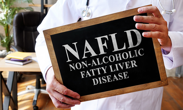
The global prevalence of NAFLD is estimated to be more that 25 percent and will continue to rise. NAFLD comprises a spectrum of liver disorders ranging from simple NAFLD to NASH. While NAFL is generally considered as to be a benign condition, NASH is prone to develop into severe end-stage liver diseases. However, the molecular mechanisms of steatosis-to-NASH progression remain poorly understood.
Using two periods of high-fat and high-cholesterol (HFHC) diet -feeding, the authors performed a large-scale integrative analysis of liver tissues from NAFL and NASH mice with their age-matched normal controls. Overall, the multi-omics study captured 176 mRNAs, 1131 lncRNAs, 48 miRNAs, 295 proteins, and 53 plasma metabolites that are altered in NAFL mice compared to the age-matched normal mice. Meanwhile, 1745 mRNAs, 5161 lncRNAs, 146 miRNAs, 674 proteins, and 82 plasma metabolites were altered in NASH mice compared to the age-matched normal mice. Through comparisons of these alterations in two mouse models, a total of 1630 mRNAs, 4547 lncRNAs, 110 miRNAs, 500 proteins, and 46 plasma metabolites were specifically altered in NASH mice compared to NAFL mice. Thus, this study provides a valuable resource to explore the molecular mechanisms of steatosis-to-NASH progression.
In addition, through transcriptomic analysis, the authors found that growth differentiation factor (GDF3) was specifically up regulated in the livers and plasma of NASH mice. In agreement, plasma GDF3 concentrations were markedly increased in patients with NASH compared to patients with NAFL or healthy individuals. Plasma GDF3 levels were strongly associated with the NAFLD activity score (NAS) and individual histologic features, including steatosis, ballooning, and lobular inflammation (Figure 1). The authors further evaluated the diagnostic potential of circulating GDF3, which demonstrated that it could be a non-invasive diagnostic biomarker for NASH patients with high accuracy.
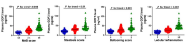
Figure One: Plasma GDF3 levels are significantly associated with histologic features in patients with NASH [Credits: Liping Xiang, Xiaoyan Li, Yunchen Luo, Bing Zhou, Yuejun Liu, Yao Li, Duojiao Wu, Lijing Jia, Pei-Wu Zhu, Ming-Hua Zheng, Hua Wang, Yan Lu].
Related topics
Animal Models, Disease Research, Genetic Analysis, Genomics, microRNA, miRNAs, RNAs