Researchers have sculpted the human body plan in a dish
Posted: 28 December 2022 | Ria Kakkad (Drug Target Review) | No comments yet
The scientists hope the human body plan will increase the understanding of the aetiology and pathology of disease.
An international team of scientists led by Dr Cantas Alev, at the Institute for the Advanced Study of Human Biology (ASHBi) at Kyoto University, Japan, have uncovered how the early stages of the human body plan are established. The study was recently published in Nature.
Like other organisms within the animal kingdom, the human body consists of repetitive anatomical units or segments – a prominent example being the vertebrae of the human spine. The most primitive version of such segments in the human embryo, known as somites, arise from an embryonic tissue called presomitic mesoderm (PSM), and contribute to the formation of various structures including cartilage, bone, skin and skeletal muscle. While previous work by Alev and colleagues reconstituted the so-called segmentation clock, a molecular oscillator and dynamic ‘wave’ of gene expression required for the proper formation of human somites (somitogenesis), it could not recapitulate the complex three-dimensional morphological and structural changes occurring during human body-axis development.
In their new study, Alev and co-workers, using a cocktail consisting of human induced pluripotent stem cells (iPSCs)-derived cells and Matrigel – a viscous gel compound enriched with extracellular matrix components – have now generated a 3D model that can recapitulate the development of our early body plan in a petri dish, which they coined axioloids.
“[Our] axioloids capture, not only the oscillatory nature of the segmentation clock, but also the molecular as well as the 3D morphological and structural characteristics observed during the process of segmentation and somitogenesis” said Alev.
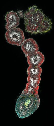
Immunofluorescence staining of human pluripotent stem cell derived axioloids
[Credit: Alev Lab (ASHBi/Kyoto University)].
By taking a bottom-up approach in their experimental design, Alev and his team identified a previously unappreciated functional role for retinoids, more commonly known as vitamin A and its derivatives, during somite formation. “Our bottom-up approach was critical to unravelling the role of retinoids during somitogenesis. It is likely that many researchers missed this essential role because vitamin A is a common supplement that usually gets included into culture media,” commented Alev.
When Alev’s axioloids were compared to actual human embryos, they revealed “remarkable similarities to Carnegie Stage 9-12 human embryos, which is known to be a critical stage during human development where organs such as the brain and heart start forming,” explained Alev.
Lastly, using iPSCs containing mutations commonly associated with congenital spine disease, Alev and co-authors demonstrated that axioloids can be instrumental in delineating how these mutations contribute to the pathogenesis of such diseases.
Alev concluded, “our [bottom-up] approach of generating axioloids have not only allowed us to uncouple fundamental biological processes, such as cell morphology and cell states, but it allowed us to determine how mutations contribute to spine disease” and he continued, “we also anticipate similar strategies will become increasingly necessary in order to understand better the aetiology and pathology of other diseases.”
Related topics
Disease Research, Genomics, Induced Pluripotent Stem Cells (iPSCs)
Related conditions
congenital spine disease
Related organisations
Kyoto University
Related people
Dr Cantas Alev