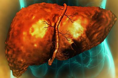
Japanese scientists have tested the material used to build models of arteries, revealing their suitability for use in medical education and surgical planning.
Hokkaido University researchers, Japan, have analysed the suitability of a smooth, flexible and transparent material used to make model arteries, for use in medical teaching and to plan for surgery on individual patients. Their work was recently described in the Journal of Vascular and Interventional Radiology.
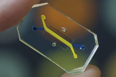
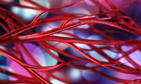
Ever-improving three-dimensional (3D) printing technology can create models of blood vessels that are significantly more realistic than those made with more conventional methods.
All types of models and simulations are applied in two key situations. They assist in medical education, allowing students of general medicine and of surgery to learn how to handle and manipulate blood vessels before applying their skills on patients. The sophistication of 3D printing also allows highly accurate models of an individual patient’s artery and vein structure to be built from radiology scans, allowing practice on a model before performing complex surgery.
The Hokkaido team has improved the assessment of the 3D printing technology by studying a material used to make model arteries that are transparent to allow careful inspection while recreating the flexibility, smoothness and slipperiness of real arteries. They observed the accuracy and suitability of the modelling materials by comparing its properties with pig arteries, measuring parameters including adhesive and tensile strength, compressibility and friction characteristics.
“We hypothesised that a tailored 3D modelling material called Flexible 80A resin would effectively simulate the properties of real arteries,” said Ryo Morita, Interventional Radiologist of the Hokkaido research group. “Our studies confirmed the material’s suitability, while at the same time identifying differences between the model material and the pig arteries that could guide further improvements.”
To avoid some limitations of this early work, moving on to use real human arteries for comparison would improve the relevance for working on patients. The methods used in this initial study could also be applied to a variety of other modelling resins to identify those most suitable.
While creating custom models for an individual patient can be of great assistance in preparing for surgery, it is expensive and time-consuming. This limits the use of personalised models to only the most anatomically challenging cases. “Making and storing models made with improved 3D printing processes could build a bank of examples for use in preoperative planning and for training,” Morita concluded.