New imaging approach reveals that changes in retinal microcirculation may indicate cerebrovascular diseases that involve reduced blood flow.
The brain is one of the most metabolically active organs in the human body. Although it represents only about 2 percent of the human body’s weight, it receives 15 to 20 percent of the body’s total blood supply. Disrupted blood flow to the brain over a long period of time, a condition known as “chronic cerebral hypoperfusion” (CCH), can lead to serious cerebrovascular diseases such as white matter disease.
CCH manifests as lesions in the white matter, a brain region vulnerable to problems with blood supply. Unfortunately, CCH has no available cure. An early diagnosis by visualising the microvascular changes in the brain that occur prior to lesion development is thus crucial. However, such a diagnosis is challenging with the available imaging techniques.
In a recent study published in Neurophotonics, researchers from USA and China led by Baoqiang Li, Associate Professor at the Chinese Academy of Sciences, investigated whether blood flow in the retina at the microscopic level could be used to predict cerebrovascular diseases involving hypoperfusion. To test this hypothesis, the team developed an innovative imaging approach based on two-photon microscopy.
Conveniently, insights into the microvasculature of the brain may come from our eyes. The retina at the back of the eye is a peripheral part of the central nervous system and shares many similarities with cerebral brain matter. But it has fewer nerve cell types and a simpler structure, making it an excellent target for studying neural circuitry and neurovascular coupling.
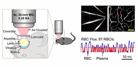
The researchers first induced CCH in mice by slightly blocking their carotid arteries. A week after, they studied one of their eyes directly under a two-photon microscope. They observed and counted red blood cells (RBCs) circulating within individual capillaries in the retinal microvasculature of mice by labelling their blood plasma with a fluorescent tag.

A new technique for retinal optical imaging developed by researchers from China and USA utilises a two-photon microscope to quantify the flux of red blood cells in microscopic capillaries within the retina, which is linked to the cerebral blood flow. (Credit: Li et al., doi 10.1117/1.NPh.10.3.035001)[/caption]
The objective of these experiments was to quantify the flux of RBCs in as many capillaries as possible. Accordingly, the researchers compared their results with those of similar measurements made on cerebral grey and white matter in a previous study performed under similar experimental conditions.
Following careful statistical analysis, they found that the mean capillary RBC flux in the retina was more significantly affected by CCH than those in the white and grey matter. While the mean RBC flux decreased by 56 percent in the retina of CCH mice compared to that in normal mice, the corresponding reductions were only 36 percent and 6 percent in their white and grey matter, respectively.
Overall, the findings of this study indicate that microcirculation in the retina could be a promising predictor of CCH, and potentially serve as an early diagnostic biomarker for cerebrovascular diseases. Moreover, the imaging approach developed by the researchers is efficient, has high signal quality, and can be implemented with a standard commercial two-photon microscope.
Neurophotonics Associate Editor Andy Shih of the Seattle Children’s Research Institute, US, remarks, “Being able to image retinal vascular physiology in vivo without adaptive optics is innovative. The results from this study may encourage further application of conventional two-photon imaging to retinal research.”