The intricate control of our bodily movements relies on a network of neural pathways connecting the brain and spinal cord. Key among these pathways is the transmission of commands from neurons in the cerebral cortex to motor neurons in the spinal cord, which in turn activate muscles, enabling fluid motion. Yet, this neural flow is disrupted in amyotrophic lateral sclerosis (ALS), a progressive neurodegenerative ailment characterised by muscle atrophy, hindering movement and breathing.
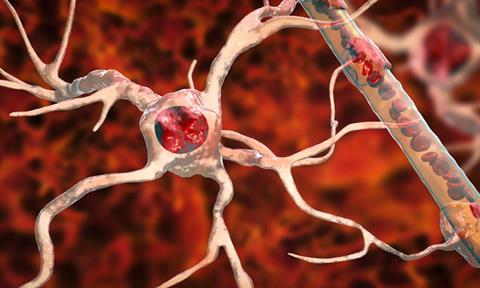
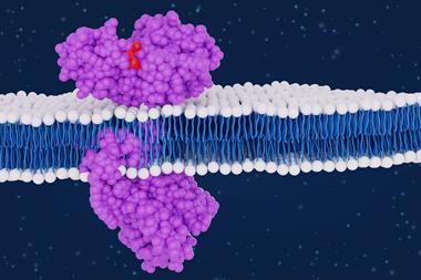
A protein named TDP-43 has been pinpointed for its abnormal accumulation in ALS-affected neurons, contributing to their decline and subsequent motor impairment.
In ALS patients, initial motor dysfunction symptoms manifest in specific body regions, like limbs, before spreading elsewhere. This pattern suggests the inception of degeneration in a specific type of motor neuron, subsequently extending to other motor-related neurons. Prior research has emphasised the presence of TDP-43 accumulation in motor neurons, coinciding with ALS. Thus, the researchers at Niigata University's Brain Research Institute pondered whether TDP-43 might be instrumental in propagating degeneration in ALS.
To dive into this query, the team engineered ALS mouse models that selectively accumulated TDP-43 in cortical motor neurons, spinal motor neurons, or skeletal muscles. Through these models, they scrutinised how TDP-43 in distinct motor neurons initiated the progression of the disease to other motor-associated neurons. Their findings were published in Acta Neuropathologica.
Dr Osamu Onodera, a professor in the Department of Neurology at Niigata University's Brain Research Institute and senior author, remarked, "TDP-43 accumulation is a common factor in most ALS patients, yet the question of whether it drives disease progression through the motor pathway has sparked long-standing debate."
The researchers discovered that TDP-43 introduced into the cortical neurons of the mouse ALS models incited mild degeneration. Additionally, they observed TDP-43 being transported along axons and conveyed to oligodendrocytes, non-neuronal cells that support neurons by enveloping axons in protective myelin sheaths, essential for efficient neuronal signal transmission.
In contrast, the induction of TDP-43 in spinal motor neurons did not disseminate to other cortical or spinal neurons, but rather triggered widespread cell death among the motor neurons and their neighbouring counterparts in the spinal cord. Furthermore, this led to profound muscle atrophy, compounding motor dysfunction.
Addressing their discoveries, co-senior author Dr Masaki Ueno, also a professor at the same institute, remarked, “Our findings suggest that pathogenic TDP-43 has multiple properties to propagate degeneration in the motor pathways in ALS, probably by spreading itself and inducing other toxic events such as degeneration and inflammation.”
The data they amassed revealed that TDP-43 diffuses across neural connections within the motor pathway, triggering various pathological responses that culminate in spinal cord degeneration. This indicates that TDP-43 employs distinct mechanisms to drive degeneration within ALS-associated motor circuits.
Dr Shintaro Tsuboguchi, the study's first author and an assistant professor at the same institute, concludes, “Elucidating the mechanisms of TDP-43 spreading and other pathological events of propagation will lead to the development of a novel therapeutic approach to prevent disease progression in ALS,” These findings hold promise for devising effective treatments for ALS, instilling hope in countless patients worldwide.