
Researchers have presented new findings that offer potential pathways to arrest critical steps toward the accumulation of mutant tau.
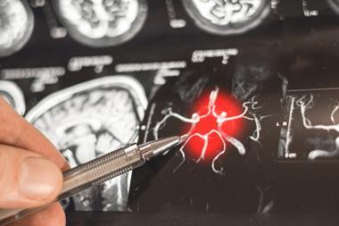
Researchers from the University of California, Santa Barbara have discovered potential ways of interrupting the misfolding of tau proteins in the brain by targeting certain sites along the long form of mutated tau. The study presents molecular-level insights on the way pathological tau spreads and could lead towards a therapeutic intervention for many neurodegenerative diseases caused by the accumulation of misfolded tau, like frontotemporal dementia (FTD), progressive supranuclear palsy (PSP) and corticobasal degeneration (CBD).
Tau is a crucial structural protein in the brain, giving cells form and stability and enabling the transport of required nutrients. However, when it mutates and misfolds, it becomes sticky and tangled. Furthermore, this folding error can become a template for faulty instructions that direct normal tau proteins to misfold and collect until the condition spreads over wide regions of the brain and interferes with brain functions. The specific locations where these neurofibrillary tangles occur in the brain differ among the neurodegenerative disorders.
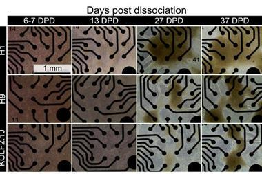
There are two forms of tau: one is produced in a short “three-repeat” version and the other is a longer “four-repeat” version. The team focused on the latter for this research. FTD, PSP and CBD are tauopathies that are far less common than Alzheimer’s disease (AD) and are exclusively of the 4R type. By using advanced techniques like transmission electron microscopy and molecular dynamics simulations with in-vitro experiments involving cell cultures, the scientists were able to explore the conditions under which pathological 4R tau begins to misfold, template other tau proteins, and aggregate.
Dr Kenneth Kosik, UCSB neuroscientist, explained: “Tau folds in a unique way in each of these diseases,” …One part of it folds into a hairpin structure only in the 4R tau.” Within the hairpin, he added, is a sticky segment called PHF6 that can bind and stack up other tau proteins into large aggregations.
He continued: “Creating conditions for tau propagation serves as a high throughput system for the discovery of compounds that may interfere with tau aggregation.” For example, they found that a single amino acid substitution along the protein adjacent to the sticky region was enough to prevent tau aggregation by weakening access to the vulnerable portion of the peptide.
The researchers conducted other investigations and discovered that nanobodies synthesised from the blood of camelids were able to bind to the PHF6 region, inhibiting aggregation of tau. The region encompassing the hairpin segment of 4R tau is the apparent active zone to target, whatever the therapy.
Although there is a long way before targeted therapeutics can be developed and approved to inhibit the formation of the neurofibrillary tangles, the results from this research offer potential pathways to arrest critical steps toward the accumulation of mutant tau.
This study is published in Proceedings of the National Academy of Sciences (PNAS).