Key function of specialised cells in peripheral nerve repair
Posted: 6 February 2019 | Iqra Farooq (Drug Target Review) | No comments yet
The interaction between macrophages and Schwann cells could be vital in maintaining the repair process, without which could impair nerve function…
Researchers have shed light on the processes behind peripheral nerve repair.
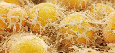
Scientists at the University of Plymouth highlighted a novel function of a cell in the immune system called a macrophage. These cells can be found in most tissues in the body. Here they are on the lookout for potential harmful organisms, and if caught, the cells destroy them. Macrophages play a role in the repair of tissues, and in the healing of wounds, following traumatic injury.
The research team identified how the macrophage uses a specific cell signalling pathway to control the processes of nerve repair following damage. Through understanding how this function works, scientists hope to be able to develop therapeutic approaches that enhance the efficacy of peripheral nerve repair.
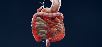
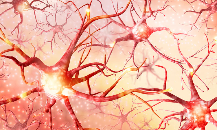
Peripheral nerves are those that connect the brain and the spinal cord to the rest of the body. Any damage to these nerves could cause a permanent loss of sensation in the area affected, or could cause an inability to control the affected muscles, or pick up and move things as we would ordinarily.
The study was conducted in mice and found that the tissue consists of several cell types, including macrophages and Schwann cells. The interaction between these cell types, and how the process allows the nerve projections to successfully regrow, is what the researchers investigated.
Macrophages were seen to form a collar around the outside of the nerve bridge and secrete high levels of a repulsive nerve guidance cue called Slit3. The also identified that Schwann cells that were inside the nerve bridge expressed the Slit3 protein Robo1.
Through these interactions, nerve projections in the bridge area regrew, which is a vital part of the repair process, without which, nerve projections would not get to their targets and could result in a permanent loss of nerve function.
Lead author Dr Xin-Peng Dun, from the Institute of Translational and Stratified Medicine (ITSMed) at the University of Plymouth explained, “Peripheral nerve injuries represent a huge clinical challenge in healthcare. More than one million people worldwide suffer from peripheral nerve damage each year – and our work has exciting potential to help patients to improve the outcome of such injuries.
“We want to understand the Slit3-Robo1 signalling and utilise it to prepare engineered nerve tissue to boost peripheral nerve repair. So it was really interesting to see the key and novel role of the macrophage, and how its production of Slit3 controls cell migration and the axons’ ‘pathfinding’ in the peripheral nerve bridge.
“We know peripheral axons have the ability to grow back into their original targets after being severed, but they do not do this quickly enough when they face the problem of long nerve gaps following injury. In this study in mice, the nerve bridges being formed were around 2mm long. But in human patients the gap can be up to 5cm to fill, so it’s important for us to keep working to explore the details of long nerve gap repair and improve the clinical management of such injuries.”
The study was published in the journal Cell Reports.
Related topics
Disease Research, Drug Discovery, Drug Targets, Research & Development, Therapeutics
Related conditions
nerve damage, Stroke
Related organisations
University of Plymouth
Related people
Dr Xin-Peng Dun