Advanced imaging technique could be used to image living cells faster
Posted: 13 December 2019 | Rachael Harper (Drug Target Review) | No comments yet
Researchers have used ghost imaging to enhance the imaging speed of nanoscopy, which could make it possible to capture processes in living cells at speeds not previously possible.
An advanced imaging technique to achieve super-resolution microscopy at unprecedented speeds and with many fewer images has been developed, which should make it possible to capture processes in living cells at speeds not previously possible and help in the development of new therapeutics.
Investigators from the Shanghai Institute of Optics and Fine Mechanics (SIOM) of the Chinese Academy of Sciences have used an unconventional imaging approach known as ghost imaging to enhance the imaging speed of nanoscopy. Their new technique produces nanometer resolution using orders of magnitude fewer images than traditional nanoscopy techniques.
“Our imaging method can potentially probe dynamics occurring on millisecond time-scales in subcellular structures with spatial resolution of tens of nanometers – the spatial and temporal resolution at which biological processes take place,” said Wang Zhongyang, co-leader of the research team.
The new approach is based on stochastic optical reconstruction microscopy (STORM), which is a wide-field technique that uses fluorescent labels that switch between light-emitting (on) and dark (off) states.
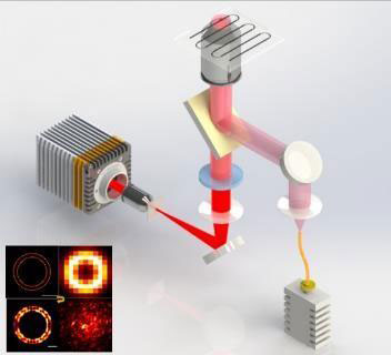
This is the nanoscopy setup used to image a 60-nm ring (inset) (credit: WANG Zhongyang).
Acquiring hundreds or thousands of images, each capturing the subset of fluorescent labels that are on at a given time, allows the location of each molecule to be determined and used to reconstruct a fluorescence image.
The researchers turned to ghost imaging to speed up the STORM imaging process. Ghost imaging forms a picture by correlating a light pattern that interacts with the object with a reference pattern that does not. The researchers also used compressive imaging, a computational approach that enables image reconstruction with fewer exposures because it uses an algorithm to fill in the missing information.
“While STORM requires a low density of fluorescent labels and many image frames, our approach can create a high-resolution image using very few frames and a high density of fluorophores,” added Han Shensheng, one of the research team’s other co-leaders. “It also doesn’t need any complex illumination, which helps reduce photobleaching and phototoxicity that could harm dynamic biological processes and living cells.”
To implement the new technique, the researchers used an optical component known as a random phase modulator to turn fluorescence from the sample into a random speckle pattern. Coding the fluorescence in this way allowed each pixel of a very fast CMOS camera to collect light intensity from the whole object in a single frame.
To form the image via ghost imaging and compressive imaging, the light intensity was correlated with a reference light pattern in a single step. The result was a more efficient image acquisition and a reduction in the number of frames required to form a high-resolution image.
The researchers tested the technique by using it to image a 60-nanometer ring. The new nanoscopy approach resolved the ring using just 10 images while traditional STORM approaches would have needed up to 4000 frames to achieve the same result. The new approach also resolved a 40-nanometer ruler with 100 images.
“We hope this method can be applied to a variety of fluorescent samples, including those that exhibit weaker fluorescence than those used in this research,” said Wang.
The researchers also said they want to make the technique faster to achieve video-rate imaging with a large field of view in order to acquire three-dimensional (3D) and colour images.
The research was published in Optica.
Related topics
Cell Cultures, Imaging, Research & Development
Related organisations
Shanghai Institute of Optics and Fine Mechanics
Related people
Han Shensheng, Wang Zhongyang