Organoids for the development of drugs and diagnostics for NASH
Posted: 3 April 2020 | Hannah Balfour (Drug Target Review) | No comments yet
Organoids at different stages of non-alcoholic steatohepatitis (NASH) progression have been created to help in drug design and identification of diagnostic biomarkers.
Researchers have created organoids that mimic the liver fibrosis characteristic of non-alcoholic steatohepatitis (NASH). The team suggest their three-dimensional (3D) cell cultures could be used to study and design drugs for different stages of NASH progression, as well as identify biomarkers for patient diagnosis.
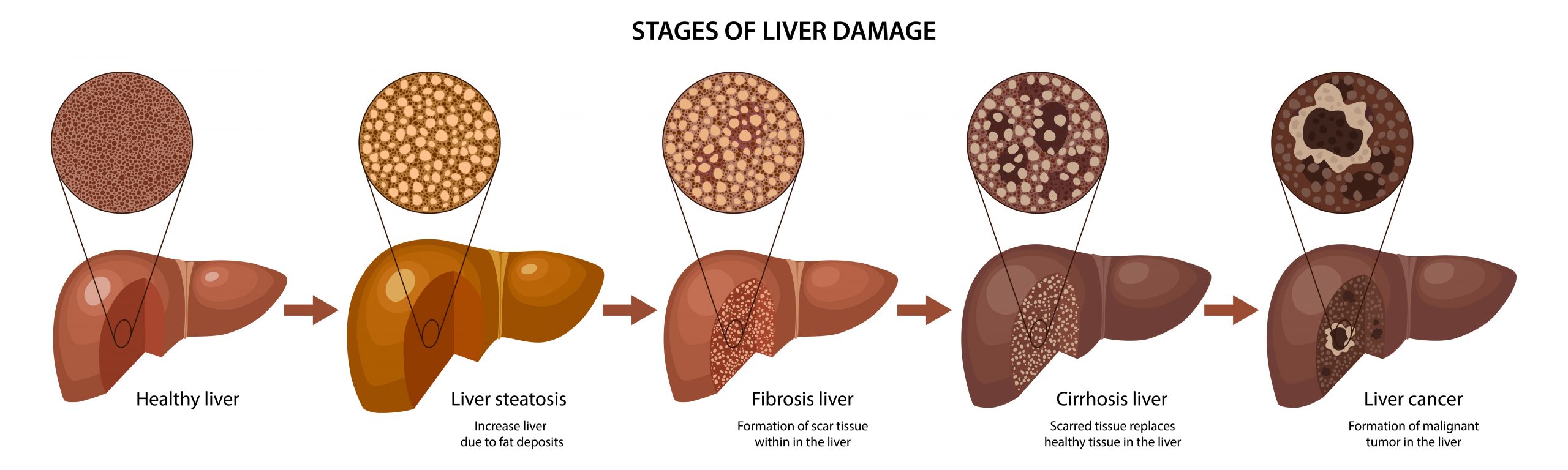
It is thought eight percent of the Japanese adult population and up to 12 percent of the adults in the US have NASH, a condition that causes patients to develop a fatty liver regardless of alcohol consumption. In more advanced stages NASH can cause liver cirrhosis and cancer. NASH symptoms include liver tissue inflammation, fat deposition and fibrosis. There are currently no medications available for treating NASH.
Traditionally drug candidates for the disease were tested on animals fed a ‘NASH-inducing diet’; however, researchers suggest this is impractical and so for their study, published in Biomaterials, they harvested cells from the livers of NASH mice at three different stages of the disease and grew them into organoids.
Biomarkers are redefining how precision therapies are discovered, validated and delivered.
This exclusive expert-led report reveals how leading teams are using biomarker science to drive faster insights, cleaner data and more targeted treatments – from discovery to diagnostics.
Inside the report:
- How leading organisations are reshaping strategy with biomarker-led approaches
- Better tools for real-time decision-making – turning complex data into faster insights
- Global standardisation and assay sensitivity – what it takes to scale across networks
Discover how biomarker science is addressing the biggest hurdles in drug discovery, translational research and precision medicine – access your free copy today
The early (fatty liver), middle (fatty liver) and advanced (advanced fibroids) stage organoids were histologically examined using stains: HE staining to visualise cell shapes, oil red staining to show oil production and Masson’s trichrome staining to highlight connective tissues. The team also used immuno-staining, quantitative PCR and RNA-sequencing to examine expression and localisation of biomarkers.
A comparison of the organoids to human NASH liver samples revealed they had similar characteristics at the three stages.
Dr Tatsuya Usui, corresponding author of the paper and Senior Assistant Professor at the Laboratory of Veterinary Pharmacology, Tokyo University of Agriculture and Technology (TUAT), Japan, concluded: “We expect that drug discovery targeted for each stage can be done using these organoids. In addition, our RNA-sequencing analysis found that several genes were elevated at all stages of organoids and some others were highly expressed at specific states (patent application filed). So far, no effective diagnostic biomarkers have been found… we expect that new reliable bio-markers for diagnosis can be identified using our NASH organoids.”
Related topics
Analysis, Analytical Techniques, Biomarkers, Cell Cultures, Disease Research, Drug Discovery, Genomics, Imaging, Organoids, Research & Development
Related conditions
Non-alcoholic steatohepatitis (NASH)
Related organisations
Tokyo University of Agriculture and Technology (TUAT)
Related people
Dr Tatsuya Usui