First 3D images of Survival Motor Neuron complex visualised
Posted: 25 March 2021 | Victoria Rees (Drug Target Review) | No comments yet
Researchers have imaged the entire Survival Motor Neuron complex using X-ray diffraction analysis, among other techniques.
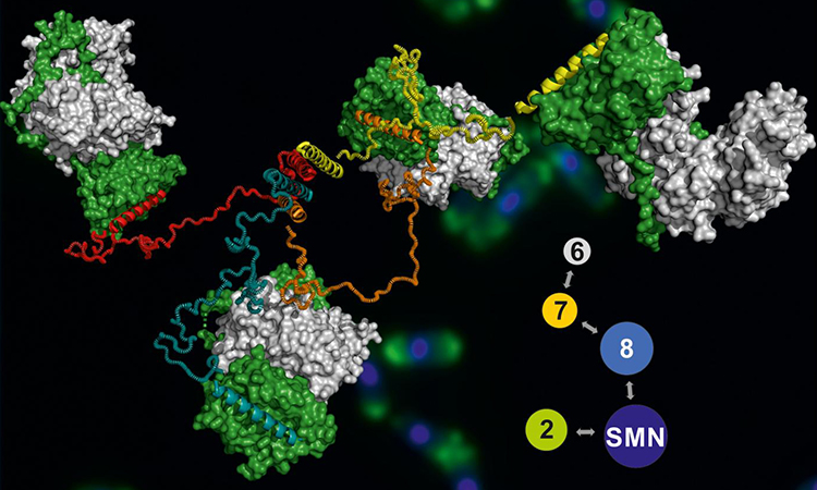
Model of the SMN complex, stained yeast cells can be seen in the background [credit: Jyotishman Veepaschit/Team Fischer].
Researchers have succeeded in presenting the first three-dimensional (3D) model of the entire Survival Motor Neuron (SNM) complex. When the SNM protein is absent, the result is the cause of spinal muscular atrophy.
Conducted at the University of Würzburg (JMU), Germany, the study now makes it possible to understand the way the complex works and why the loss of its function leads to muscular atrophy.
“The structural analysis of large and complex molecules in atomic detail has been made possible by the ‘revolution-resolution’, which was primarily brought about by the developments in cryo-electron microscopy,” said Professor Utz Fischer, lead researcher from the study.
Biomarkers aren’t just supporting drug discovery – they’re driving it
FREE market report
From smarter trials to faster insights, this report unpacks the science, strategy and real-world impact behind the next generation of precision therapies.
What you’ll unlock:
- How biomarkers are guiding dose selection and early efficacy decisions in complex trials
- Why multi-omics, liquid biopsy and digital tools are redefining the discovery process
- What makes lab data regulatory-ready and why alignment matters from day one
Explore how biomarkers are shaping early drug development
Access the full report – it’s free!
However, according to the team, the challenge with this technology is that it works best on structures that are more or less rigid and have few flexible sections. Unfortunately, many molecular entities are not built like this, including the SMN complex.
“This complex is of central importance for our cells because it supports the formation of molecular machines required for the expression of our genes,” said Fischer. To serve its function in the cell, it must be highly flexible and dynamic. As a result, structural analysis by traditional strategies has been impossible so far. Therefore the Fischer and his team chose an alternative approach.
“Our starting point was a co-operation with the group of Dr Rémy Bordonné from Montpellier in France, that enabled us to identify the SMN complex of the yeast Schizosaccharomyces pombe,” he explained. This complex was ideally suited for an integrative structural analysis because it comprises less individual components than its human counterpart and has a less dynamic behaviour.
“In a first step, we visualised, by X-ray diffraction analysis, individual sections that are important for keeping the complex together,” said Fischer. In a second step, they characterised the entire complex and parts of it by means of small-angle X-ray scattering. This method also provides information on the dynamic behaviour of unfolded sections of the complex. In parallel, missing sections were reconstructed by means of a bioinformatic method called 3D homology modelling.
The result was a model of the entire SMN complex that provides an excellent explanation of its function. The team found that the central “body” of the complex has a number of long and very flexible “arms” which enable the complex to catch proteins and assemble them, together with other biomolecules, into molecular machines.
The model also provides insight into the processes that lead to spinal muscular atrophy. The mutations causing this disease are concentrated in the central body, the researchers say. They prevent the full development of the complex, so that it cannot serve its function in the cell.
The study is published in Nucleic Acids Research.
Related topics
Imaging, Informatics, Microscopy, Protein, Proteomics, Structural Biology
Related conditions
Spinal muscular atrophy
Related organisations
University of Würzburg (JMU)
Related people
Professor Utz Fischer