New imaging method visualises respiratory activity of cell models
Posted: 3 May 2021 | Victoria Rees (Drug Target Review) | No comments yet
Researchers have created a new imaging technique called electrochemiluminescence to visualise multiple spheroids with a single shot.
Researchers from Tohoku University, Japan, have shone a light on the process that cells use to exchange gases with their environment by developing a new technique called electrochemiluminescence (ECL) imaging.
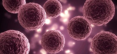
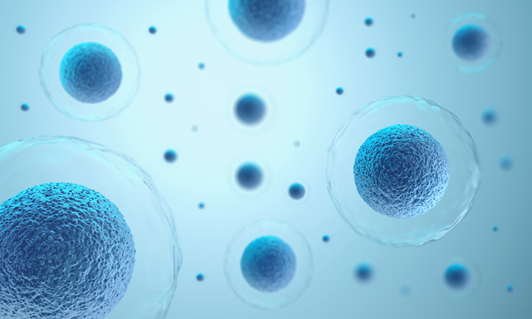
The team used spheroids – cultured cells within a close-to-natural environment – to mimic a biological tissue using mesenchymal stem cells (MSCs). Due to MCSs’ ability to self-renew and differentiate into various tissues, they are of significant interest for use in regenerative medicine and to test different therapeutics in different tissue models.
They settled the spheroids on a gold electrode dosed in luminescent solution. The researchers applied stepped electric potential – the energy needed to convert oxygen – to the set up where the solution was then oxidised sensitised to produce luminescent, which is dependent on oxygen concentration. The spheroids glowed brightly, surrounded by a ring of darkness where oxygen concentration was low due to gas exchanges. They call the process potential step-based ECL imaging.
“As living spheroids consume oxygen to produce energy, the respiratory activity was elegantly visualised by the distribution of luminescence around the spheroids,” said Kaoru Hiramoto, who developed the ECL imaging system.
The researchers used a digital camera to visualise multiple spheroids with a single shot, demonstrating both the high spatial resolution of their system, as well as the low background noise.
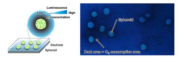
Spheroids were settled on the gold electrode with a luminescent solution. When the potential steps were applied, the spheroids were brightly visualised. The dark area around the spheroids indicates a decrease in luminescence attributed to low O2 concentration, caused by the respiratory activity of the spheroids [credit: Kaoru Hiramoto, et al.].
“The system offers high-throughput analysis of spheroids, in addition to highly improving resolution of the images compared to conventional electrode array devices,” said co-corresponding authors Associate Professor Kosuke Ino and Professor Hitoshi Shiku, researchers on the study. “The system offers novel insights into electrochemical devices and imaging systems for cell spheroids.”
The team plan to improve the sensitivity and selectivity of the system, as well as analyse other data that might be collected in the system, such as cellular metabolites attached to the electrode. They also plan to use the system to investigate more complicated biological models, as well as patient-derived cell spheroids.
“To the best of our knowledge, this is the first attempt to visualise the respiratory activity of spheroids by direct conversion of oxygen concentration into ECL imaging,” Ino said. “The features of the proposed system – high spatial resolution with the ability for simultaneous imaging of multiple spheroids – are promising for transplantation research and drug screening utilising cell spheroids.”
The study was published in Biosensors and Bioelectronics.
Related topics
Disease Research, Imaging, Label-Free, Regenerative Medicine, Stem Cells
Related organisations
Tohoku University
Related people
Associate Professor Kosuke Ino, Kaoru Hiramoto, Professor Hitoshi Shiku