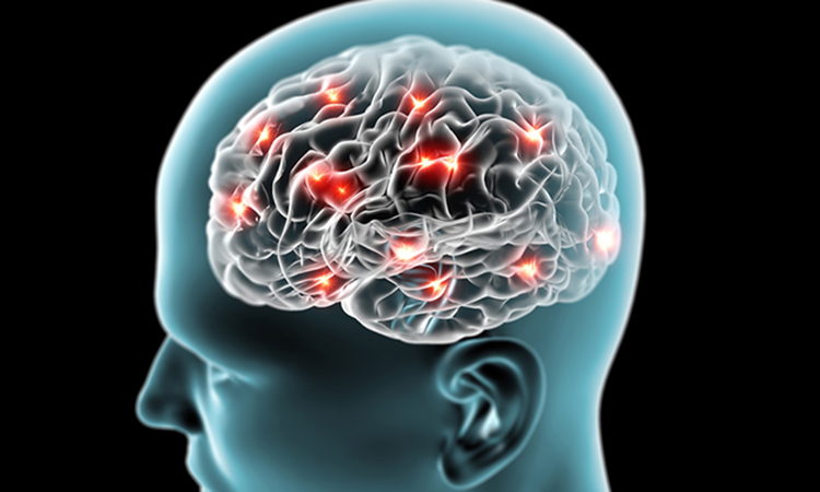
Study provides clues to cause and possible treatment of Parkinson’s
Posted: 14 June 2021 | Victoria Rees (Drug Target Review) | No comments yet
Researchers have shown that the leakage of mitochondrial double-stranded DNA into the cell can contribute to Parkinson’s disease.
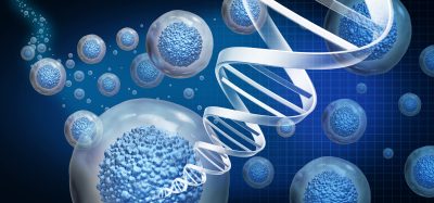
Researchers from Niigata University, Japan, have developed a new approach that could revolutionise the treatment, prevention and possibly reversal of the damages that lead to Parkinson’s disease. This novel finding, in cellular and zebrafish models, demonstrates how the leakage of mitochondrial double-stranded DNA (dsDNA) into the cytosol environment of the cell can contribute to the impairment of brain tissue of patients with Parkinson’s disease.
“Our results showed for the first time that cytosolic dsDNA of mitochondrial origin leaking and escaping from lysosomal degradation can induce cytotoxicity both in cultured cells, as well as in zebrafish models of Parkinson’s disease,” said lead researcher Professor Hideaki Matsui. “This study showed that the leakage of this mitochondrial nucleic material may occur as a result of mitochondrial dysfunction, which may involve genetic mutations in genes encoding mitochondrial proteins or incomplete degradation of mitochondrial dsDNA in the lysosome – which is a “degradation factory” of the cell.”
According to the researchers, upon the leakage into the cytoplasm, this undegraded dsDNA is detected by a “foreign” DNA sensor of the cytoplasm (IFI16) which then triggers the upregulation of mRNAs encoding for inflammatory proteins (type I interferon stimulated cytokines such as IL1β). The team hypothesise that the subsequent accumulation of inflammatory protein within the cytoplasm may cause cell functional imbalance and ultimately cell death.
“However, this dsDNA leakage effect can be counteracted by DNAse II, a dsDNA degrading agent,” said Professor Akiyoshi Kakita, an associate investigator in the study.
The first part of the study was conducted in vitro, using cells of nerve cancer origin (SH-SY5Y cells) with defective mitochondria and lysosomal dysfunctions through knockdown of GBA, ATP13A and PINK1 genes. The mutant cells demonstrated leakage of dsDNA and accumulation of inflammatory cytokines and cell death. In an additional comparison experiment using mutant cells (with defective mitochondrial proteins) and wild type SH-SY5Y cells, they further demonstrated that DNAse II rescued cells through the degradation of dsDNA.
In a confirmatory study using a Parkinson’s zebrafish model (gba mutant), the researchers demonstrated that a combination of Parkinson’s-like phenotypes including accumulation of cytosol dsDNA deposits, reduced the number of dopaminergic neurons after three months. Lastly, they further generated a DNase II mutant zebrafish model which exhibited decreased numbers of dopaminergic neurons and demonstrated accumulated cytosolic DNA. When the original zebrafish was complemented with human DNAse II gene, the overexpression of human DNAse II decreased cytosolic dsDNA deposits and rescued neurodegradation by saving the number of dopaminergic and noradrenergic neurons after three months.
The researchers say this demonstrated that neurodegenerative phenotype of gba mutant zebrafish induced by dsDNA deposits in the cytosol can be restored by DNAse II.
To determine the effect of cytosolic dsDNA of mitochondrial origin in human brain with Parkinson’s, they inspected postmortem human brain tissues from patients with idiopathic Parkinson’s. They observed an abundance of cytosolic dsDNA of mitochondrial origin in medulla oblongata of postmortem brain tissues, the levels of IFI16 were also markedly increased in these brain tissues. Taken together, results in this study demonstrated that cytosolic dsDNA of mitochondrial origin accumulated in PD brains and that these dsDNA deposits and IFI16 play contributory roles in human PD pathogenesis.
The study was published in Nature Communications.
Related topics
DNA, In Vitro, In Vivo, Molecular Targets, Neurosciences
Related conditions
Parkinson's disease
Related organisations
Niigata University
Related people
Professor Akiyoshi Kakita, Professor Hideaki Matsui