ProteinSimple Peggy Sue

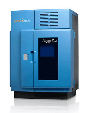
Simple Western™ system that can separate, analyse and quantify proteins based on either size or charge…
Two ways to get separation, identification and quantitation
Peggy Sue™ lets you separate and analyzse proteins by size or charge from 2-440 kDa either by immunoassay or total protein. Got small sample volumes or starting materials? No problem. She uses as little as 0.2 µg/µL of protein in just 5 µL of sample, and runs up to 96 samples in one experiment. Charge ahead and do more with Peggy Sue!
TECHNOLOGY & APPLICATIONS
Peggy Sue provides two different perspectives for quantitating biological responses.
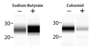
FIGURE 1. Analyzing post-translational modifications of Histone H3 is no problem for Peggy Sue. Left: HeLa cell lysate from cells stimulated with sodium butyrate was probed with anti-acetyl Histone H3 (Lys9) to characterize changes in acetylation. Right: Methylation was analysed in lysate from HeLa cells stimulated with colcemid and probed with anti-dimethyl Histone H3 (Lys39).
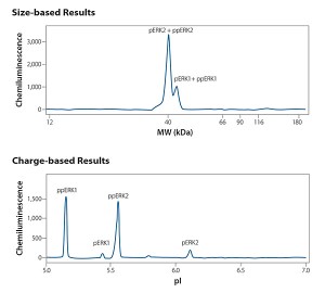
FIGURE 2. The same HeLa lysate sample, stimulated with epidermal growth factor, was analysed and probed with an anti-phospho specific ERK antibody in both size-based and charge-based assays. The size-based electropherogram shows detection of total phosphorylated ERK2 (pERK2 + ppERK2), a quantifiable large peak; and total phosphorylated ERK1 (pERK1 + ppERK1), a smaller peak, at their correct molecular weights. The charge-based electropherogram provides more in-depth characterization of the single and dual phosphorylated isoforms of ERK1 and ERK2 at their expected pIs, demonstrating that the dual phosphorylated isoforms are the dominant species present in the lysate.
Downloads
Learn More
Click here to find out more about the ProteinSimple Peggy Sue
Alternatively, click here to request a quote
Related content from this organisation
Related topics
Assays, Cell-based assays, Drug Targets, Screening, Translational Science
Related organisations
ProteinSimple