


whitepaper
ebook: Optimise your high value cell lines
As interest in biotherapeutic proteins grows, the need to reduce cell line development costs and the time to market is more critical than ever.
List view / Grid view
As interest in biotherapeutic proteins grows, the need to reduce cell line development costs and the time to market is more critical than ever.
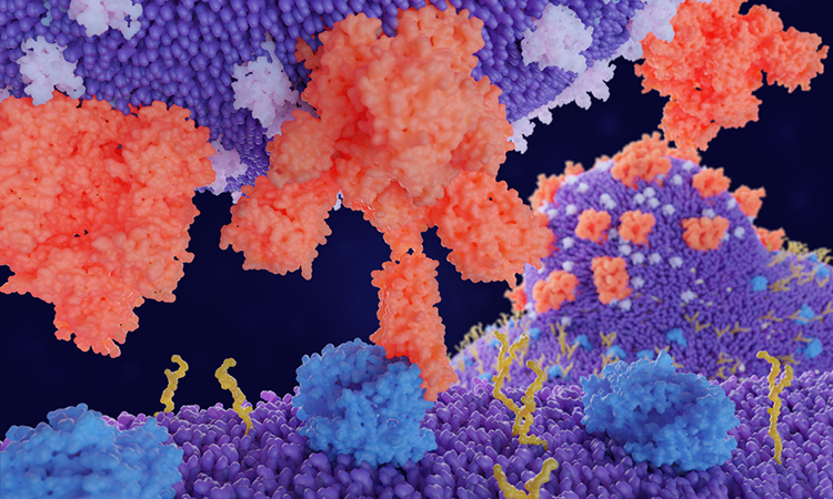
A study has uncovered previously unknown properties of the Spike protein from the SARS-CoV-2 Alpha and Beta variants.

Solutions to help you understand viral diseases and translate your research findings into better treatments and vaccines.

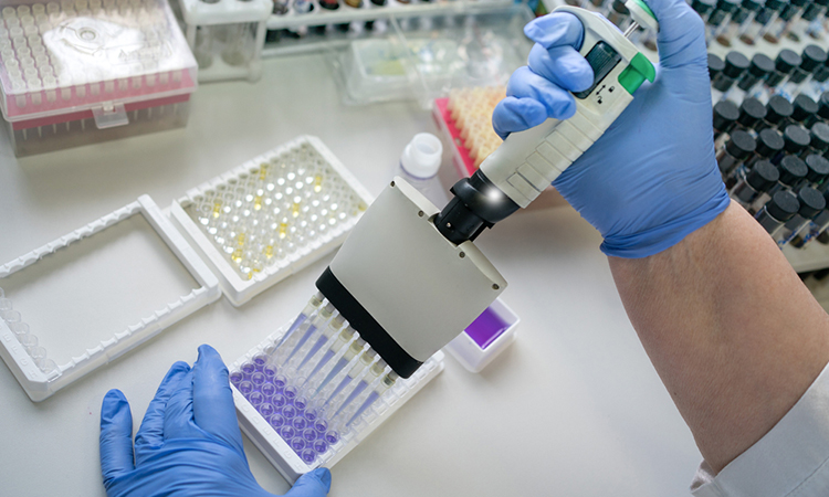
While researchers conduct their studies, constraints such as time can impact their work. Dr Ian Holland from the University of Edinburgh spoke with Drug Target Review’s Deputy Editor Victoria Rees to explain how lab automation can offer a solution to these challenges and enhance output for scientists.
Scientists have used imaging methods and machine learning to understand cellular metabolism at the single-cell level.

Despite many companies considering digital transformation a top priority, research shows that 70% of digital transformation initiatives fail. In this ebook, we explore why and how the right approach can see your organisation succeed.

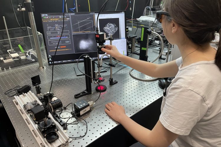
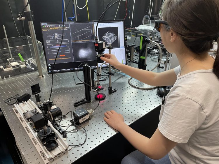
Researchers have created an X-ray scanning machine that shows the shape of an object and its molecular composition.

Complete solutions for neurological disease research and discovery - helping you to better understand diseases to improve patient outcomes.
A new non-invasive microscopic fluorescence imaging method has been developed to reveal details of the brain in animal models of various diseases.
A modified luciferase enzyme has been developed as an immunosensor to be fused with a Q-body and used in immunoassays.
A wide range of tools to support your immuno-oncology research and help redefine and develop tailored, life-changing immunotherapies to fight cancer.
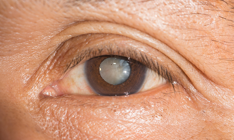
Researchers have shown that a protein named aquaporin can disrupt optical development, leading to cataract formation.
Our comprehensive portfolio suite is designed to optimize your productivity to get safe and efficacious vaccines and therapeutics to market faster.
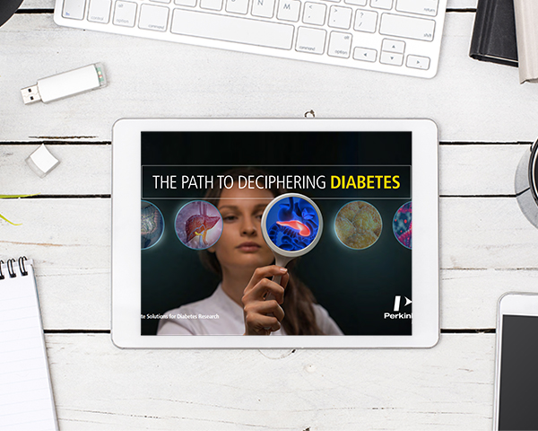
Solutions to aid understanding of cellular and molecular pathways in diabetes and translate these findings into prevention and treatment strategies.
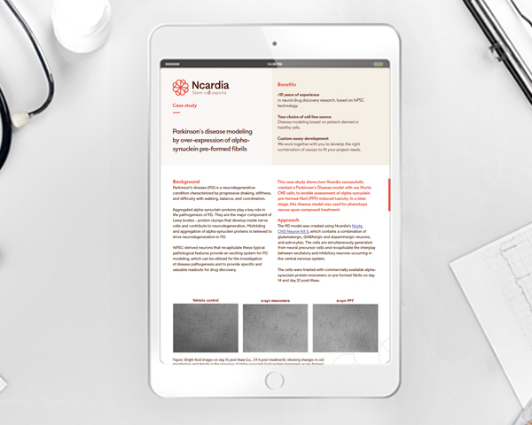
The development of a Parkinson’s disease model, using hiPSC-derived neural cells, to assess alpha-synuclein pre-formed fibril-induced toxicity.