Expert view: New developments in real‑time live‑cell analysis
Posted: 5 September 2018 | Del Trezise | No comments yet
Live-cell analysis is now a well-established method that is part of the everyday cell biologist’s armoury for understanding cell biology. For over a decade, the functionality, throughput, and ease of use offered by real-time live-cell analysis has provided a platform for accurate and reproducible cell biology analysis in multiple biomedical research areas, including oncology, immuno-oncology, immunology and now neuroscience.
With ever‑expanding datasets, real‑time live‑cell analysis is evolving to remain a tool that researchers need in order to create physiologically relevant cellular models.
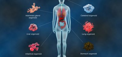
More advanced cell models place a higher demand or expectation on the analytical technique. In oncology and immuno‑oncology, for instance, 3D cell culture models represent a more relevant biological model. With the right combination of culture plate and, if required, extracellular matrix material, real‑time live‑cell analysis platforms can support advanced 3D spheroid cell culture models to allow simple and cost‑effective growth and health assays of cancer cells.
Another important step in expanding our ability to create accurate cell models, is defining individual cellular heterogeneity within the context of cellular populations. New image processing algorithms have been developed for real‑time live‑cell analysis systems that allow label‑free cell counting, observations of basic cell morphology at the single‑cell level, and intracellular fluorescence intensity measurements.
In addition, methods are now available to allow immunocytochemistry on living cells by labelling surface proteins with fluorescent antibody fragments. With this labelling method and time‑lapse imaging, it is possible to observe patterns of expression over time and track cells interacting through extracellular protein binding.
These methods represent only a few of the advancements made in real‑time live‑cell analysis recently but demonstrate how the method is continually evolving to remain an integral tool in the cell biologist’s toolbox. Coupled with other more traditional approaches, such as flow cytometry and high definition imaging, real-time live‑cell analysis will continue to provide insights toward advanced, physiologically relevant cell modelling.
Related topics
Antibodies, Assays, Cell Cultures, Flow Cytometry, Imaging, Immuno-oncology, Immunology, Neurosciences, Oncology, Protein, Research & Development
Related organisations
Essen BioScience
Related people
Del Trezise