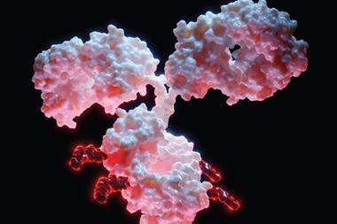
A single change in the amino acid structure of the transmembrane segment can either enhance or diminish the inhibitory function of PD-1 in immune responses.
Researchers from NYU’s Langone Health’s Perlmutter Cancer Center and the University of Oxford have shown how treatments to restrict PD-1’s action could be strengthened to improve their anticancer effect. The study also supports experimental treatment strategies for autoimmune diseases, as stimulating PD-1 action could block an overactive immune response.
One of the most important checkpoints is the programmed cell death receptor (PD-1) protein, which is blocked by checkpoint inhibitors, a fairly new drug class. The study authors state that these drugs are somewhat effective in a third of patients with a variety of cancers, but the field is urgently exploring ways to improve their performance and scope.
As well as this, PD-1 signalling is slowed in autoimmune diseases such as rheumatoid arthritis, lupus, and type 1 diabetes, meaning that the action of unchecked immune cells creates inflammation that damages tissues. Drugs that stimulate PD-1, named agonists, are currently showing promise in clinical trials.
Numerous immune checkpoints are receptors on the surface of T cells. These translate docking information from the outside of the cell to the signalling portion of the receptor inside the cell. The transmembrane segment connects the outside-of-the-cell portion of PD-1 with the inside portion. Although many immune receptors function in pairs called dimers, until now, it was believed that PD-1 functioned alone, not in the dimer form.
However, the new study found that PD-1 forms a dimer through interactions of its transmembrane segment. This finding is in sharp contrast to other immune receptors, which typically form dimers through the segment of the receptor that is outside the cell. Immune cell testing in mice demonstrated that encouraging PD-1 to form dimers, specifically in the transmembrane domain but not in its outer or inner regions, increased its ability to suppress T cell activity. Decreasing transmembrane dimerization lowered PD-1’s ability to inhibit immune cell activity.
Dr Elliot Philips, study lead investigator and internal medicine resident at NYU Grossman School of Medicine and Perlmutter Cancer Center explained: “Our study reveals that the PD-1 receptor functions optimally as dimers driven by interactions within the transmembrane domain on the surface of T cells, contrary to the dogma that PD-1 is a monomer.”
Co-senior investigator and structural biologist Dr Xiang-Peng Kong, added: “Our goal is to use our new knowledge of the functioning of PD-1 to determine if weakening its dimerization, or pairing, helps make anticancer immunotherapies more effective, and just as importantly, to see if strengthening its dimerization helps in the design of agonist drugs that quiet overactive T cells, tamping down the inflammation seen in autoimmune diseases.”
It was also discovered that a single change in the amino acid structure of the transmembrane segment can either enhance or diminish the inhibitory function of PD-1 in immune responses. The team plans further investigations of PD-1 inhibitors and agonists to see if they can design more effective therapies for both cancer and autoimmune disorders.
This study was published in Science Immunology.